Quantcast
Channel: Special Section – Lower Extremity Review Magazine
Viewing all 184 articles
Browse latest View live

Hip disorders in children with Down syndrome

$
0
0
surestep-1

Photo courtesy of SureStep.

As hip arthritis becomes a growing concern in adults with Down syndrome, clinicians are also becoming more attuned to hip issues in children with DS in hopes that early intervention in the pediatric population will help reduce the risk of disability later on.

By Shalmali Pal

Based on the numbers alone, hip disorders in children with Down syndrome (DS) don’t seem to be a cause for great concern—it’s estimated that only 5% to 8% of this patient population will suffer from hip impairments, with the most common being subluxation.1 But the detrimental effects of hip disorders can have serious implications, most notably the development of early arthritis.

“Fortunately, [hip disorders] in children with Down’s are relatively uncommon. But when it’s bad, it’s bad, so you don’t want to outright dismiss it [as a possibility],” said Stephen England, MD, a pediatric orthopedic surgeon at Park Nicolette Health Partners in St. Louis, MN.

Orthopedic problems are the second most common cause of disability (after cardiac issues) in adult patients with DS, who are living more active, longer lives (See “Down syndrome and total hip arthroplasty: Opportunities to optimize outcomes,” LER March 2015, page 16).

“Now that cardiac issues, such as coronary artery disease, are being identified and treated more aggressively, and because the lifespan of children with DS is longer, these [orthopedic] problems will definitely be more of an issue for the adult DS population,” said Stephen Sundberg, MD, a pediatric orthopedic surgeon and program medical director of the Center for Pediatric Orthopedics at Gillette Children’s Specialty Healthcare in St. Paul, MN.

LER spoke with Sundberg, England, and other biomechanics experts about the diagnosis and management of hip disorders in children with DS.

The goal of physical therapy in children with Down syndrome is to “lengthen and strengthen” the muscles so that the hip remains centered in the socket.
Photo courtesy of SureStep.

Photo courtesy of SureStep.

Common causes

There are three main reasons for hip disorders in kids with DS. One of the least prevalent is Legg-Calve-Perthes (LCP) disease, where the head of the femur loses blood supply and the bone becomes weak and misshapen. This condition generally manifests in children aged 4 to 8 years as a painless limp and the loss of full range of motion in the hip.1 LCP occurs in children with and without DS, but data on how commonly it’s seen in the latter population are scant, according to England.

Then there is slipped capital femoral epiphysis—also known as epiphysiolysis, but generally called SCFE (pronounced “skiffee”)—in which the rounded head of the femur slides on the femoral neck. This hip impairment is linked with obesity and hypothyroidism. SCFE presents as a limp with associated pain in the hip or even the knee.1

“SCFE generally hits right around puberty,” explained Kathy Martin, PT, DHS, a professor and director of the Doctor of Physical Therapy Program at the Krannert School of Physical Therapy, University of Indianapolis. “Because this disorder is closely tied with the onset of puberty, we feel like there is some hormonal element involved. So the increased load from being overweight plus hormonal changes may put some kids with DS at a greater risk for SCFE.”

Finally, there is subluxation, where the head of the femur moves out of the acetabulum.

“This dislocation may or may not be associated with malformation of the acetabulum. The dislocation appears to be due to a combination of laxity of the connective tissue that normally keeps the hip together along with the low muscle tone found in DS,” wrote pediatrician Len Leshin, MD, in a 2003 online article.1

Sundberg described subluxation as a deficiency in the posterior wall of the hip socket in children with DS.

“We think that the back wall of the socket is relatively underdeveloped, and that can predispose a small percentage of these kids to develop a progressive hip dislocation,” he said.

England added that subluxation can be painless, with the hip joint popping in and out of place.

“You or I would no doubt feel that, but for a child with DS, that ability to pop the hip out may become something they are accustomed to,” England said.

Sundberg emphasized that practitioners will need input from caregivers and parents to make the correct diagnosis.

“We generally find that kids with DS may not tell us if something is painful,” he said. “We have to rely on the families to give us the feedback. They’ll tell us that, ‘My child is behaving different. He used to be able to join us for family walks or was willing to come with me to the grocery store. Now he wants to stay at home and spends more time sitting.’”

Martin also stressed obtaining feedback from parents or other caregivers who know the child best. Just as parents can tell whether an infant’s cry means “I’m hungry” or “I’m tired,” they can offer insight into why a child is refusing a routine activity.

“They can often discern some of the subtle behaviors that may indicate a physical problem or may just mean, ‘I don’t want to do this today,’” she said.

Subluxation can manifest at any age. Ideally, pediatricians are assessing for potential hip dysplasia during wellness check-ups in all newborns and infants, the experts agreed. Accepted physical tests for dysplasia include the Barlow and Ortolani tests.2

Martin said subluxation is something she sees more often in infants and toddlers than in older children.

“Many orthopedic surgeons consider a congenitally dislocated hip as an orthopedic emergency. It needs to be addressed and treated immediately,” she said, or patients face poor outcomes, such as the early onset of hip osteoarthritis.3

If a hip disorder is suspected, the child can be sent for an orthopedic exam that may include x-rays. But England cautioned that a static x-ray may show a completely normal hip, as subluxation is a dynamic condition.

Arthrograms will reveal subluxation more readily, allowing clinicians to look specifically at the dynamic relationship between the femoral head and the cup, England said, “but arthrograms are not something you want to do routinely because they require anesthesia.”

He emphasized that before children with DS undergo any invasive procedure requiring anesthesia, they need to be checked for laxity in the cervical joints (C1/C2) as instability in those locations could lead to “potentially catastrophic” problems if the neck is hyperextended to accommodate intubation. These problems can include deterioration of gait, loss of manipulative skills, and hemiparesis.4

Sundberg added that assessing hip health in children with DS may require more vigilant observation than in a typical child. Hip subluxation goes through stages; a child may begin life with normal hips but can develop progressive subluxation over the course of many years, he said.

“The hip may be moving in and out of the socket, and the child may not complain of pain early on,” he said. “But they may ultimately go on to develop a fixed dislocation where the hip no longer wants to go back into the socket. That’s not a situation we want to leave unaddressed.”

The surgical approach

Are all children with DS and hip disorders candidates for surgery? There are numerous factors that go into making that call.

A 2006 review article5 on the role of the orthopedic surgeon in DS offered clear cut-advice: “In the absence of radiographic changes in a child whose hip dislocates, we recommend immobilization in a hip spica cast or hip abduction orthosis. When radiographic changes are evident in the acetabulum or when signs of subluxation of the hip are present, we recommend surgical intervention.”

However, Sundberg said he is not convinced that casting or bracing will keep patients out of the surgical suite. He explained that even with some level of success with nonsurgical methods to reduce subluxation, children with DS will still have progressive migration or shallowness of the hip socket.

“They are more than likely to end up needing surgery,” he said.

A Bernese periacetabular osteotomy (PAO) is the preferred surgical intervention to reorient the socket, Sundberg said. Studies have shown good success rates with PAO, with patients maintaining high Harris Hip scores at six months and one year postsurgery, as well as clinical stability and asymptomatic improvement at five years.6,7

Sundberg said his group has done the procedure in children with DS who were as young as 8 years.

“There’s an age range that depends upon when the instability develops,” he said. “If the kids are younger and have no open growth plates, then we may do the PAO. If subluxation becomes a problem as the kids approach skeletal maturity, ages twelve to fourteen, we may consider a standard, adult-style osteotomy.”

Caregivers need to be aware that surgical adjustment of subluxation in childhood is no guarantee that the patients won’t experience hip disorders in adulthood, England pointed out.

“The goal with the surgery is to prevent that joint from popping in and out, but the joint is still likely to wear out, which can lead to arthritis,”3 he said. “I don’t necessarily think [surgery] leads to long-term joint preservation; I think it’s more about short-term joint stability.”

Lengthen and strengthen

As with surgery, conservative treatment options for kids with DS and hip disorders depend on the type of impairment and the child’s age.

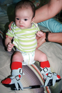
For infants with subluxation, the standard in conservative therapy is the Pavlik harness, which limits range of motion of the hip joint. Unlike a rigid cast, the harness allows the child to move freely within a certain range.

“It allows kids to flex and externally rotate, but it limits their ability to go into adduction,” Martin said. “It keeps them out of the end range of the position where the hip is likely to pop out.”

But the Pavlik harness is most successful in children aged up to 6 months. In a Turkish study of harness use for developmental hip dysplasia (DHH) in children with a mean age of 8 weeks, 18 of 31 procedures were deemed successful with no avascular necrosis at follow-up.8 A UK study of 123 hips treated in an infant DHH screening program found that all hips diagnosed with dysplasia or subluxation, but not dislocation, were managed successfully in the harness.9

If the subluxation isn’t discovered until the child is older, then a closed reduction under general anesthesia is likely necessary. The child is then put in a spica cast to keep the hip in position during healing.2,5

Photo courtesy of SureStep.

Photo courtesy of SureStep.

When it comes to getting the most out of noninvasive treatments, Martin said, “the earlier, the better,” as the development of the acetabulum is limited after the first 18 months of life.

“If the joint is still developing and remodeling, you can guide how it remodels and make it more stable in that first eighteen months,” she said. “If you’ve missed that window of opportunity, then you are limited as to what changes you can make in that joint.”

Those with LCP and SCFE are more likely to be candidates for surgery. In the case of LCP, the procedure will establish proper alignment of the hip bones. In the case of SCFE, surgery is a must as it will speed up the epiphysis closure.10,11

Of course, physical therapy (PT) is paramount for all patients, experts say, regardless of whether they’ve undergone correction with surgery, bracing, or casting.

The goal with PT in this group is not so much to impact the joints directly, but to “lengthen and strengthen” the muscles around the hip, balancing the muscles so that the hip remains centered in the socket. Martin pointed out that children with DS have what she called “a double whammy.”

“They have low muscle tone, and they have ligamentous laxity. The ligaments are the primary joint stabilizers, so if those aren’t working, then you rely on secondary joint stabilizers to protect the joints, and those are the muscles,” she said. “We are trying to make sure that there is optimal range of motion, and then maximizing the strength of the muscles to help support the hip.”

Feet first

The hypotonia or hypermobility that predisposes children with DS to hip disorders also is often manifested in the foot.12,13 Making the connection between better foot health and better hip health in the DS population has been a shortcoming in the medical community, according to Louis DeCaro, DPM, of the DeCaro Total Foot Care Center in West Hatfield, MA, who is also president of the American College of Foot and Ankle Pediatrics.

“I think that there are certainly many specialists that don’t consider the significant impact of the foot on short- or long-term hip health. I think that they see a lot of [kids with DS] where they make judgment calls about the hips, but don’t realize that the foundation has not been addressed well,” he said.

Martin agreed that addressing foot impairments in children with DS may often go unexamined, especially by general medicine practitioners.

“I think [addressing the feet] is becoming more common, with the idea of providing support as soon as kids with DS are showing interest in getting on their feet, but it’s PTs and developmental pediatricians who are initiating that,” she said.

Research showing causation between foot laxity and hip instability is lacking, but connections between ligamentous laxity and musculoskeletal disorders have been demonstrated. A 2004 study looked at 125 children who presented at a pediatric rheumatology clinic with painful conditions related to hypermobility. The authors reported that the majority of the patients had pes planus and muscle weakness. Of the total population, 12% had what the authors called “clicky” hips at birth, and 4% had a congenital dislocatable hip.13 A 2013 article from Gillette pointed out that “excessive ankle joint laxity and pes planus are more common in patients who have joint hypermobility.”14

“I think about alignment of the whole lower extremity,” Martin said. “If you help align the foot, then everything stacks up better. [For] a child with a collapsed arch or pronated foot—it’s like driving a car with the front end out of alignment. The car will move, but there’s going to be abnormal wear and tear.”

Treatment options for foot problems in children with DS include flexible supramalleolar orthoses (SMO) or custom foot ankle orthoses.

In children with DS and hypotonia (mean age approximately 6 years), Martin reported significant improvement with SMOs versus shoes only for the standing, walking, running, and jumping dimensions of the Gross Motor Function Measure. Results were seen immediately and after seven weeks of use.15

In another study in children with DS aged 3 to 6 years, wearing custom foot orthoses was associated with decreased ankle eversion during static standing, and various improvements during walking, such as a decreased foot progression angle and a change in the initial contact site from flat foot to heel strike.16

The overall goal with orthoses is to improve coordination, balance, posture, and stability during gait, DeCaro said.

For instance, for the typically flexible DS foot, DeCaro said he has found that a functional UCBL (University of California Biomechanics Laboratory) type of orthosis combined with supportive footwear can be effective. He believes all orthoses for children with DS should have a deep heel cup, a medial heel skive, and high medial and lateral sidewall flanges.9

Asked if addressing alignment problems from the feet up in these young patients could potentially prevent hip impairments as they grow older, he replied: “Unequivocally yes! A good foundation is imperative for a lifelong improvement of overall gross motor strength and biomechanics.”

Shalmali Pal is a freelance writer based in Tucson, AZ.

REFERENCES

1. Leshin L. Musculoskeletal Disorders in Down Syndrome website. http://www.ds-health.com/ortho.htm. Accessed May 4, 2015.

2. Hip dysplasia. Stanford University School of Medicine website. http://newborns.stanford.edu/HipDysplasia.html#management. Accessed May 4, 2015.

3. Jackson JC, Runge MM, Nye NS. Common questions about developmental dysplasia of the hip. Am Fam Physician 2014;90(12):843-850.

4. Charleton P, Dennis J, Ellis J, et al. Cervical spine disorders: craniovertebral instability. Down Syndrome Medical Interest Group (DSMIG UK). Published March 2012. http://www.dsmig.org.uk/library/articles/CSI%20revision%20final%202012.pdf

5. Caird MS, Wills BPD, Dormans JP. Down syndrome in children: the role of the orthopaedic surgeon. J Am Acad Orthop Surg 2006;14(11):610-619.

6. De la Rocha A, Sucato DJ, Tulchin K, et al. Treatment of adolescents with a periacetabular osteotomy after previous pelvic surgery. Clin Orthop Relat Res 2012;470(9):2583-2590.

7. Katz DA, Kim YJ, Millis MB. Periacetabular osteotomy in patients with Down’s syndrome. J Bone Joint Surg Br 2005;87(4):544-547.

8. Atalar H, Sayli U, Yavuz OY, et al. Indicators of successful use of the Pavlik harness in infants with developmental dysplasia of the hip. Int Orthop 2007;31(2):145-150.

9. Walton MJ, Isaacson Z, McMillan D, et al. The success of management with the Pavlik harness for developmental dysplasia of the hip using a United Kingdom screening programme and ultrasound-guided supervision. J Bone Joint Surg Br 2010;92(7):1013-1016.

10. Pinheiro PC. Nonoperative treatment of slipped capital femoral epiphysis: a scientific study. J Orthop Surg Res 2011;6:10.

11. A patient’s guide to slipped capital femoral epiphysis. OrthoPediatrics website. http://www.orthopediatrics.com/docs/Guides/slipped_CFE.html. Accessed May 4, 2015.

12. Aprin H, Zink WP, Hall JE. Management of dislocation of the hip in Down syndrome. J Pediatr Orthop 1985;5(4):428-431.

13. Adib N, Davies K, Grahame R, et al. Joint hypermobility syndrome in childhood. A not so benign multisystem disorder? Rheumatology 2005;44(6):744-750.

14. Quanbeck D, Greer K, Wilkins K. Joint hypermobility: normal variation or cause of concern? Gillette Children’s Specialty Healthcare: A Pediatric Perspective 2013;22(1):1-3.

15. Martin K. Effects of supramalleolar orthoses on postural stability in children with Down syndrome Dev Med Child Neuro 2004;46(6):406-411.

16. Selby-Silverstein L, Hillstrom HJ, Palisano RJ. The effect of foot orthoses on standing foot posture and gait of young children with Down syndrome. NeuroRehabilitation 2001;16(3):183-193.

Kinematics identify subgroups of kids with CP and equinovarus

$
0
0
prokinetics-1

Photo courtesy of ProtoKinetics.

Treatment planning could benefit

By Barbara Boughton

Chicago researchers have identified clinically relevant subgroups based on foot and ankle kinematics in children with equinovarus secondary to hemiplegic cerebral palsy (CP) that could help improve treatment planning and clinical outcomes.

The researchers used gait analysis and multisegment foot and ankle modeling to classify 24 children with hemiplegic CP, all of whom had a clinical diagnosis of unilateral equinovarus, and 20 typically developing children into five subgroups.

“There is a lot of variability in characteristic foot deformities among children with equinovarus secondary to hemiplegic cerebral palsy,” said lead author Joseph Krzak, PT, PhD, PCS, assistant professor of physical therapy at Midwestern University in Downers Grove, IL, and senior motion analysis laboratory physical therapist at the Shriners Hospital for Children in Chicago. “As a result of this variability, as well as multiple combinations of surgeries used to correct equinovarus, there is a lot of variability in treatment outcomes.”

In fact, postoperative success rates range from 67% to 82% for surgeries aimed at correcting equinovarus in hemiplegic CP, the researchers noted in their paper, which was published in the February issue of Gait & Posture.

“Hopefully our research could be used to improve outcomes, since children with similar foot deformities are likely to benefit from similar surgical interventions,” Krzak said.

The researchers used the Milwaukee Foot Model, which employs radiographic skeletal indexing, to measure and quantify the 3D kinematics of small foot segments during locomotion. Five gait classifications for the children in the study were developed using principal component analysis and cluster analysis. One subgroup of 15 typically developing children and three children with CP, all of whom had a rectus foot type with a well-aligned hindfoot and forefoot, served as a control group. Four other subgroups were classified according to the presence or absence of equinovarus deformities: those with just hindfoot involvement, both hindfoot and forefoot involvement, varus deformity with hindfoot and forefoot involvement but no equinus, and forefoot adductus but no hindfoot varus or equinus.

The gait classifications developed in the study could be used to plan surgical and nonsurgical interventions.

In the subgroup without equinus or varus, the chief complaint for patients with CP was the in-toeing often seen in that patient population—which can come from internal rotation of the trunk, pelvis, hip, lower leg, hindfoot, and/or forefoot, Krzak said.

“Although equinovarus deformity may have contributed to a chief complaint of in-toeing, the deformity at the foot and ankle may have been only a small contributor,” he said.

Eventually, the gait classifications developed in the study could be used, not only for treatment planning, but also to evaluate the outcomes of surgery.

“Right now the assessment of surgical outcomes is observational,” Krzak said. “But, if the subgroup classifications we develop are validated in future studies, they could be used to quantitatively measure the outcomes of surgery.”

The next step for the researchers is to validate their findings in a larger group of 100 children with equinovarus secondary to hemiplegic CP; that study is currently being planned, Krzak said.

Krzak acknowledged that the technology used in the study was quite sophisticated and not available to most practitioners.

“One future direction of our research could be to show how clinical tools that most clinicians have access to could be used to classify and assess children based on the subgroups we identified,” he said.

The study by Krzak and colleagues provides insight into the variability of foot deformities that exist in equinovarus secondary to hemiplegic CP, said Susan Rethlefsen, PT, DPT, a physical therapist at the motion analysis laboratory at the Children’s Orthopaedic Center in Los Angeles, whose work centers on assessing children with CP who are candidates for surgery.

“The study’s findings do verify what I see clinically in terms of the different types of deformities,” Rethlefsen said.

It is difficult to look at an equinovarus foot and assess just where the deformity arises, she acknowledged.

“This study takes us one step closer to being able to measure that accurately,” Rethlefsen said.

Although most of the deformities in the study were fairly severe—since all the children with CP were candidates for surgery—the subgroup classifications could also be used for less severe cases and for nonsurgical treatment planning, Krzak said.

Rethlefsen concurred.

“Knowing the characteristics of a foot deformity could help an orthotist position the foot correctly during casting,” she said.

Barbara Boughton is a freelance medical writer based in the San Francisco Bay Area.

Source:

Krzak JJ, Corcos DM, Damiano DL, et al. Kinematic foot types in youth with equinovarus secondary to hemiplegia. Gait Posture 2015;41(2):402-408.

Lower-body focus could help youth baseball player arms

$
0
0

5PEDS-news-baseball-iStock14153096-copyStudies link hip, shoulder function

By Chris Klingenberg

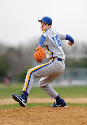
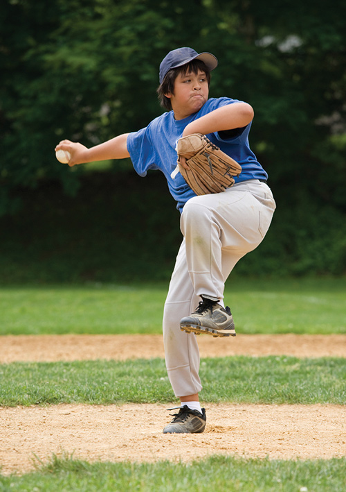
Kids grow up thinking that throwing a baseball hard is all about strength in the arm and shoulder, but new evidence suggests that muscle strength and range of motion in the hip affect shoulder function during throwing in youth baseball players.

Gretchen Oliver, PhD, FACSM, ATC, an assistant professor in the School of Kinesiology at Auburn University in Alabama, and colleagues found that rotational passive range of motion (PROM) in the throwing shoulder was significantly correlated with hip PROM in 26 youth baseball pitchers with no history of injury.

“A pitcher needs adequate stance leg internal rotation for the windup, then as they drive off the rubber they need stance hip external rotation. Just as the stride hip needs adequate external rotation to get the best foot contact, once the ball is released, they need to have adequate stride hip internal rotation for the body and arm to decelerate around the stride hip,” Oliver said.

A single examiner measured bilateral hip and throwing shoulder rotational PROM in the youth pitchers using an inclinometer. The players averaged 31.3° of internal rotation and 35° of external rotation in the stance hip and 28.5° of internal rotation and 37.2° of external rotation in the lead hip. In the throwing shoulder, mean internal rotation was 34.5° and external rotation was 110.5°. The differences between internal and external rotation were statistically significant for the shoulder and lead hip, but total PROM did not differ significantly between the stance and lead hips. The findings were epublished in December by the Journal of Strength and Conditioning Research.

“Internal and external rotation of the hips should average forty-five degrees. Deviations from that are going to ultimately result in problems,” Oliver said.

Michael Hannon, MD, an orthopedic surgeon at the Kerlan-Jobe Orthopedic Clinic in Los Angeles, and his colleagues have also assessed both upper and lower extremity function in preadolescent and adolescent baseball players.

“Ultimately, the most important thing with younger kids is that people focus so much on arm strength when it is really the lower extremity that we should be focusing on,” Hannon said.

“If the core and lower extremities are weak, there is a better chance of throwing out your arm.”
— Michael Hannon, MD

The Kerlan-Jobe study analyzed 54 preadolescents (mean age 9.9 years, 38 pitchers) and 54 adolescents (mean age 15.1 years; 34 pitchers) with regard to hip ROM, hip abduction strength, scapular dyskinesis, coracoid distance, and single-leg squat testing. All players were male. There was a clear difference in the number of years pitched: a mean of 2.9 years in the preadolescent group compared with 8.9 in the adolescent group. The adolescent pitchers were throwing an average of 9.2 months per year, whereas the preadolescents averaged only 5.7 months per year.

There was a significant difference in hip internal rotation between the preadolescent group and the adolescent group for the dominant or stance leg (40.81° vs 33.09°) and the nondominant or stride leg (38.37° vs 34.35°). External rotation did not differ significantly between the two age groups for either leg. There was a significant difference in dominant hip ROM (78.46° vs 71.63°) between the preadolescent and adolescent groups. No difference was seen when considering the total hip ROM of the nondominant hip. The findings were published in August 2014 by the American Journal of Sports Medicine.

For the single-leg squat test, participants were first asked to stand on one leg and establish single-leg balance, then squat to at least 60° and return to the baseline position. The test was considered positive if the player was unable to maintain single-leg balance, was unable to maintain level hips, or exhibited significant hip adduction or internal rotation during squatting. None of the 54 preadolescents could complete the single-leg squat test on either leg; in the adolescent group, 13% performed the test successfully on the stance leg and 9% were successful on the stride leg.

“It is important that coaches are informed that the core and lower extremity are critical to avoid arm problems,” Hannon said. “If the core and lower extremities are weak, there is a better chance of throwing out your arm.”

Oliver underscored the benefits of the single-leg squat test for examining balance and pelvic stability.

“Can they balance on one leg and, if they can, do they have the gluteus medius strength to stabilize their pelvis to perform the squat? Usually after a few tries, they get the balance down, and then we see a corkscrew from the hip down as they do not have the glute-med strength to stabilize the pelvis, which then results in knee valgus,” she said.

Chris Klingenberg is a freelance writer based in Massachusetts.

Sources:

Oliver GD, Weimar WH. Hip and shoulder range of motion in youth baseball players. J Strength Cond Res 2014 Dec 5. [Epub ahead of print]

Beckett M, Hannon M, Ropiak C, et al. Clinical assessment of scapula and hip joint function in preadolescent and adolescent baseball players. Am J Sports Med 2014;42(10):2502-2509

Obese children develop knee malalignment as they mature

$
0
0

5PEDS-news-obesity-iStock29518762-copyEffect might increase future OA risk

By Emily Delzell

As obese children undergo the rapid physical changes of puberty, they develop knee malalignment that could potentially contribute to development of knee osteoarthritis (OA), according to recent research from Nationwide Children’s Hospital in Columbus, OH.

“Children start in varus and, typically, around age seven or eight, progress to normal alignment. Our study found that obese children begin with slightly higher varus [than nonobese children] that moves to greater valgus alignment in late puberty,” said study lead author Sharon Bout-Tabaku, MD, assistant professor of pediatrics at Nationwide Children’s.

She and colleagues pooled data from three groups of children and adolescents: healthy participants aged 4 to 17 years; children aged 4 to 17 years taking part in a weight management program; and obese adolescents aged 13 to 17 years enrolled in a clinical trial of the weight loss drug sibutramine.

The investigators stratified participants into nonobese and obese groups based on body mass index z-scores (BMI-Z), or standard deviations, from a reference population. They defined obesity as a BMI Z-score at or above the 95th percentile for the reference standard and ranked pubertal age by Tanner stage. They assessed body composition and knee alignment with DEXA (dual-energy x-ray absorptiometry), measuring metaphyseal-diaphyseal angle (MDA) and anterior tibiofemoral angle (ATFA).

Obese children begin with greater varus than nonobese children, which shifts to greater valgus alignment in late puberty.

More than half (52%) of the 320 participants were obese. Although obese children were older overall (11.9 vs 10.5 years) obese participants at Tanner stages 3 and 4 were significantly younger than their nonobese counterparts.

Mean ATFA and MDA angles didn’t differ according to obesity status, and neither angle was correlated with BMI-Z score or fat mass Z-scores. MDA values, however, did vary by Tanner stage and obesity status.

Obese Tanner stage 1 participants had significantly greater MDA, indicating less valgus alignment, while obese Tanner stage 4 and 5 adolescents had significantly lower MDA, compared with nonobese children. Sex-stratified analyses showed heavier girls had greater variability in knee alignment than those weighing less.

The Journal of Rheumatology published the study in January.

“It may be that the extra weight children carry puts more pressure on developing joints so they don’t straighten out as they grow, but we need to follow children longitudinally to see whether excess weight actually changes the way knees and bones develop,” Bout-Tabaku said.

Sarah Shultz, PhD, ATC, a lecturer in biomechanics at the School of Sport and Exercise at Massey University in Wellington, Australia, has studied dynamic alignment in obese prepubertal children, finding they often use a more valgus gait to prevent additional loading on the medial compartment of the knee.

“This difference could mean needing a wider stance, or trying to shift forces more laterally through the knee,” she said. “Regardless, the fact that prepubertal children are choosing this pattern supports the idea that a valgus gait promotes abnormal bone growth due to abnormal mechanical loading, and could lead to a more permanent static alignment in pubertal obese children.”

Whether—and how early—this increases risk of OA in children is still unknown, said Bout-Tabaku, but a Austrian study epublished in May 2014 by Knee Surgery, Sports Traumatology, Arthroscopy did identify signs of early OA in extremely heavy teenagers.

Magnetic resonance imaging scans of 39 morbidly obese adolescents with and without knee pain revealed that all but one had a marked cartilage lesion in at least one knee region, and the group reporting pain had significantly more lesions.

The next step for Bout-Tabaku is seeing whether losing weight restores normal alignment, which she and her colleagues will look at in an upcoming study.

She pointed out that the major limitation of her current study was the use of supine DEXA scans rather than standing x-rays to measure knee alignment and that had children been weight-bearing, the significant differences between the obese and nonobese groups likely would have been amplified.

Shultz, who in her own research has used DEXA to measure ATFA, noted its use might be why, in contradiction with other studies of static alignment, the current investigation didn’t find greater valgus alignment in younger obese children.

Bout-Tabaku agreed DEXA needs further validation as a measure of knee alignment, but sees potential value for the modality.

“DEXA exposes children to less radiation and allows us to look at adiposity, which causes inflammation that may be causative for OA,” she said.

Sources:

Bout-Tabaku S, Shults J, Zemel BS, et al. Obesity is associated with greater valgus knee alignment in pubertal children, and higher body mass index is associated with greater variability in knee alignment in girls. J Rheumatol 2015;42(1):126-133.

Shultz SP, D’Hondt E, Fink PW, et al. The effects of pediatric obesity on dynamic joint malalignment during gait. Clin Biomech 2014;29(7):835-838.

Widhalm HK, Seemann R, Hamboeck M, et al. Osteoarthritis in morbidly obese children and adolescents, an age-matched controlled study. Knee Surg Sports Traumatol Arthrosc 2014 May 20. [Epub ahead of print]

Orthotic devices and CMT: Everyone deserves an aha! moment

$
0
0

Susan-AcropolisThis guide to orthotic devices for people with Charcot-Marie-Tooth (CMT) disease has been a dream of mine for a long time. I was diagnosed with CMT1A at the tender age of 18 months, but didn’t learn about orthotic devices for CMT until 2007, when I started a support group in Atlanta. Up to that point, I thought ankle foot orthoses (AFOs) were for the aged and infirm—something that, at age 35, I didn’t think I was.

When orthotist Sean McKale, CO, LO, came to my support group meeting, he immediately recognized in me the walk of someone with CMT. When Sean, who sits on the CMT Association (CMTA) Advisory Board, shared this with me, I was shocked. I had no difficulty walking—or so I thought! However, I listened closely as he talked about the benefits of in-shoe foot orthoses to correct alignment and prevent joint deterioration. My mother had just had a double knee replacement, necessitated by poor alignment caused by her CMT. OK, I thought, maybe I could use foot orthoses to prevent that type of joint deterioration.

As Sean went on to detail the benefits of AFOs, especially the dynamic AFOs, I got progressively more excited. Did I want to fall less? Yes. Did I want more energy throughout my day? Yes! Did I want to stop reduce my joint pain, especially in my knees, hips, and back? YES! OK, Sean, I thought, I’ll try a pair of AFOs.

That was my aha! moment. The AFOs felt like very lightweight, stable boots with springs in the heels. They looked like shin guards, the kind used in soccer. They could be easily camouflaged under pants, with the right shoes. Yes, I thought, maybe I could use these from time to time.

Six years later, I wear them about 50% of the time. I love to hike in the woods, where my AFOs prevent trips and falls. I love to travel, and my AFOs prevent fatigue from long walks and rolling my ankles on cobblestones. Overall, my AFOs allow me to walk further, longer, and to have less pain and more energy at the end of an arduous day. I can do the things I love to do!

I have not become dependent upon my AFOs. I don’t wear them when I know I won’t walk far or when I would prefer to wear shorts or a dress. I don’t wear them to exercise on the bicycle or treadmill, where I can hold on for balance. I have learned when I can go without them and when I can’t. I have worked hard to maintain strength and muscle tone in my legs. My AFOs supplement my leg strength rather than replacing it, vastly improving my quality of life.

This guide is aimed at helping others to reach their aha! moments faster than I did.

Together with Lower Extremity Review and the extensive input of CMTA Advisory Board members Sean McKale and David Misener, CPO, we hope this publication will empower patients with CMT and practitioners to discuss different types of orthotic devices and make an informed decision about which type will be the best fit.

The CMTA’s vision is a world without CMT. We will achieve this vision through our extensive and focused drug development research initiative, STAR (Strategy to Accelerate Research). We know that we will soon be successful in finding treatments to slow, stop, and reverse the progression of CMT.

Until then, we believe it is our responsibility to provide resources to people living with CMT about treatments that can improve quality of life, and, when indicated, those include in-shoe foot orthoses and AFOs.

I encourage you to visit our website, cmtausa.org, for research updates, educational resources, to connect with others in the CMT community, or to invest in our vision of a world without CMT by making a donation. And please feel free to call us at 800/606-2682 or email us at info@cmtausa.org.

Wishing you the best of health,

Susan Ruediger
Director of Development
Charcot-Marie-Tooth Association

Jeff Seitzer: AFOs and staying fit help manage his mild symptoms

$
0
0

Jeff-Seitzer-1By Robyn Parets

It’s hard to imagine that a 56-year-old man who swims, plays golf, works out, and even practices yoga has Charcot-Marie-Tooth disease (CMT). But Jeff Seitzer, who was diagnosed more than 40 years ago, has managed to keep his symptoms at bay. He believes staying fit and flexible has been the key to maintaining his active lifestyle.

In fact, Seitzer says he noticed an increase in his stamina and energy level when he started attending yoga classes about 10 years ago. At home, he also stretches “maniacally,” and has noticed that his legs stiffen up when he doesn’t both stretch and exercise regularly.

Seitzer was about 13 years old when he was diagnosed with CMTX, which is typically associated with more severe symptoms in men than women due to its linkage to the X chromosome. Clinically, CMTX is similar to the more prevalent CMT1 subtype; common symptoms include muscle weakness and atrophy, and loss of feeling in the feet, lower legs, and hands.

Seitzer only has mild symptoms, which include foot drop, weakness, and pain in his legs and feet at times. Even his doctors at the CMT Clinic at the University of Iowa in Iowa City can’t believe how much he can still do.

Jeff-Seitzer-2“By my age, I should be hobbled,” Seitzer said.

Born in Omaha, NE, Seitzer now lives in Chicago with his wife, Janet Smith, and 8-year-old daughter, Penelope (who is adopted and therefore not at risk of inheriting CMT from Seitzer). The University of Chicago alum teaches religion and philosophy at Roosevelt University in downtown Chicago and often rides his bicycle to work. To help ease pain and extend his stride when walking, Seitzer has been wearing Kinetic Research’s Noodle AFOs (ankle foot orthoses) since 2014. Before that, he tried other types of orthotic devices, but he says the Noodle AFOs are a major step up for him. For example, they are particularly useful when wearing winter boots to walk on ice and in snow, as they help to improve his stability.

“They are a game changer,” he said.

Seitzer also said the Noodle offers support during other activities, like riding his bike. For instance, he said, when wearing the AFOs he can ride his bike without needing to stretch first. Also, the Noodle’s carbon fiber foot plate almost acts as a “biking shoe,” helping distribute his weight more evenly on the pedals.

If Seitzer had his Noodle AFOs 40 years ago, they might have saved him some embarrassment, too. That’s because, as a teenager, he wore metal braces. At the time, Seitzer’s mother, who also had CMT, thought she was doing the best thing for him. She didn’t want him to end up like her father, who had a severe case of the disorder. As a result, Seitzer wore the awkward braces until he was about 20.

Jeff-Seitzer-4“They were not so great for those teenage years,” he recalled.

Seitzer’s mother was also hyper-vigilant about curbing the progression of her son’s CMT for another reason: At the age of four, he suffered from a major bout of encephalitis, which left him temporarily paralyzed on his right side. Other residual side effects left Seitzer with extreme nervous energy, memory loss, and mood swings. He also had reconstructive surgery on his right foot, which was malaligned and weakened by the encephalitis.

“It was devastating,” he said.

Despite a difficult childhood and his share of struggles, Seitzer considers himself lucky to be leading an active adult life.

“I have good and bad days,” he said. “I understand my body, and it’s an ongoing process.”

Robyn Parets is a freelance writer in the Boston area.

Photos by Joshua Albanese (joshuaalbanese.com).

Brennan Ryan: With AFOs and cool sneakers, this 10-year-old can be a regular kid

$
0
0

Brennan-RyanBy Robyn Parets

If you ask Brennan Ryan what his favorite after-school activities are, he’ll tell you, “Basketball, baseball, flag football, and Cub scouts.” Ask him what his favorite part of school is and he’ll answer, “Recess.” This isn’t surprising considering that Brennan Ryan is 10 years old and in fourth grade.

“He’s a regular boy. He likes sports and has a ton of friends,” said Kathy Ryan, Brennan’s mother.

But, at the same time, Brennan, who has Charcot-Marie-Tooth disease (CMT, specifically subtype CMT1A), is no ordinary kid. His mother, who has extremely mild CMT symptoms, knew something was wrong from the time her son was six months old. He had trouble moving around and didn’t start walking until he was around two years old. The developmentally delayed baby started physical therapy before his first birthday and, as a toddler, he had trouble with balance and couldn’t run or jump like the other kids, Kathy said.

Once diagnosed with CMT through genetic testing, Brennan immediately began wearing SureStep SMOs (supramalleolar orthoses) to help his excessive pronation and other symptoms.

“The SMOs helped his overall stability and gait. They also gave him confidence,” Kathy said.

Brennan-2Orthotic devices have played a bigger role in Brennan’s life as his CMT symptoms have become more severe over the years, said Kathy, who also has twin 12-year-old daughters who do not have CMT. Brennan becomes fatigued easily, frequently has pain in his feet and hands, and has trouble with weakness in his extremities. For example, his thumb dislocates often and he has difficulty holding a pencil. In school he uses technology, including an iPad with a keyboard, to help him write and keep up in class, said Kathy, who is a third and fourth grade teacher. Perhaps the hardest thing is watching her son come home from school wiped out or just lie on the floor after playing sports.

“It takes him a while to gain his energy back,” she said,

Brennan goes to physical therapy once a week, and his mom also helps him stretch his legs at home. They’ve found that it’s important for him to do these exercises regularly, Kathy said.

“He’s a very bright boy and very aware of what he has [to deal with],” she said.

However, Brennan is happy to have orthotic devices that allow him to wear cool sneakers, just like his friends. Kathy and Brennan’s father, Kevin Ryan, just have to search a little harder for the extra-wide footwear brands that will fit over the devices.

“He wants the two-hundred dollar gym shoes that the other kids have,” Kathy joked.

Fitting in socially is certainly as important as shoes that fit, at least if you’re a kid from suburban Chicago like Brennan. At the same time, the Ryan family believes education about CMT and other neuromuscular disorders helps children accept differences among their peers. To this end, Brennan was happy to be chosen last year as a Muscular Dystrophy Association (MDA) Goodwill Ambassador for the state of Illinois.

Brennan-Ryan-3“It was pretty cool. I got to do a photo shoot with a [Chicago] Bears player,” Brennan said.

Kathy explained that her usually more reserved son came out of his shell to appear on MDA posters and marketing materials. He also made some public appearances. One of these included going to an elementary school to talk to the kids about CMT and other neuromuscular diseases.

“I liked it, and I think I helped other kids,” Brennan said.

Robyn Parets is a freelance writer in the Boston area.

Photos by Joshua Albanese (joshuaalbanese.com).

Diane Covington: AFOs positively impact every aspect of her dynamic life

$
0
0

Diane-profileBy Jordana Bieze Foster

It’s a happy coincidence that Diane Covington lives just a few miles from a New Balance store, for two reasons. The brand’s sporty and comfort styles are perfectly suited for Covington’s life on the go. And they come in widths that can accommodate the ankle foot orthoses (AFOs) that make her active lifestyle possible.

Covington, 59, was diagnosed with Charcot-Marie-Tooth (CMT) disease 30 years ago, after coworkers’ comments about her limping gait prompted her to see a neurologist. Later, when blood testing for CMT had become more widely available, her subtype was identified as CMT2A—and, in retrospect, she believes her father and paternal grandmother likely had CMT as well. The oldest of Covington’s three adult children has also been clinically diagnosed with CMT.

Covington’s symptoms include impaired balance, muscle weakness, and diminished sensation in her extremities. But, like many people with CMT, Covington was reluctant to try AFOs until her symptoms had deteriorated to the point at which she could not stand unassisted and became fatigued after 15 minutes of shopping at the mall. That was nine years ago. Now, she wears her Custom Composite AFOs literally from when she wakes up until she goes to sleep (except when bathing), and she wishes she had embraced the idea earlier.

Diane-2“If I had to say anything to people with CMT about AFOs, I would say, ‘Don’t wait,’” Covington said. “We think it’s going to be this devastating experience, but in fact it’s just the opposite. There is nothing about my life that my AFOs haven’t impacted in a positive way. I think they’re just the best.”

The stability and support provided by the AFOs allows Covington to pursue a number of outdoor activities near her home outside of Boise, ID, including Nordic walking, riding a recumbent tricycle, and exploring the local landscape in an ultra-terrain vehicle with her fiancé, Mike Steiner. But she says some of the smaller achievements have made the biggest difference.

“I can now stand up by myself in the middle of a room,” Covington said. “That doesn’t sound like a big feat, but before I would have had to hold onto something in order to stand. I really would not be able to leave the house safely without them.”

To be sure, finding the right pair of AFOs has been a process. Covington estimates she has tried 10 different pairs of AFOs and seen at least three orthotists, and not just because she and Steiner relocated to Idaho from Colorado last year. Some designs were uncomfortable, some were not durable enough, and some were too wide to fit in any conventional footwear. It took about six years to finally find her AFO soulmates.

“I would also tell people with CMT to not be shy about changing orthotists if the two of you are not on the same wavelength,” Covington said. “Find someone who understands CMT, because not everyone does.”

Diane-3Over time, Covington has become more comfortable disclosing her condition to others—something that was particularly challenging when she reentered the dating world several years ago as a divorcee. Steiner has convinced her that in hot weather it makes sense to wear shorts, but typically she still prefers long pants over skirts or capris and lace-up shoes over sandals for better AFO camouflage.

“I can verbalize about the AFOs, but showing them off is another thing,” she said.

In addition to her lower extremity issues, Covington also has diminished sensation in her fingers, but that hasn’t stopped her from typing, quilting, and creating glass mosaics.

“I’m not at the point where I feel deprived because I have CMT,” she said. “I’m just grateful that I’m almost sixty and it really hasn’t impacted my life too much.”

Story and Photos By Jordana Bieze Foster


SureStep: A Revolutionary Bracing Concept

$
0
0

surestep-2AFO Mechanics

SureStep manufactures a variety of products but is primarily known for the SMO (Supra malleolar orthosis) and pediatric AFOs (ankle foot orthosis). The SureStep SMO is meant for patients up to 80 pounds with hypotonic presentations. The purpose is to control the side to side movements of the ankle. The trim lines can be extended longer laterally or medially depending on whether the patient is pronating or supinating. As patients grow older there is a device called the Big Shot that accommodate children and adults weighing over 80 pounds. What makes the SureStep SMO so unique is the dynamic function that the brace allows. It uses compression to create alignment, unique trim-lines to allow freedom of motion where needed, and made from a material that travels together along with the foot.

Controling the Tri-Planar Deformities

SureStep BigShot

SureStep BigShot

This works under compression to maintain stability through midline. In young patients with CMT they can possibly present with a more over pronated foot position because of the low muscle tone (hypotonia) prior to developing a high arch foot presentation. This can been seen in CMT patients who have some symptoms of the disease but have not developed a high arch otherwise known as pes cavus foot. The SMO holds the heel in vertical alignment, giving stability in a coronal plane (side to side) position. An SMO is not necessarily for a patient who has weaknesses pushing up or down the ankle, just poor side-to-side movement.

Improving Stability During Gait

The device is designed to allow for motion to still occur from in and out of the midline position but limits the extremes that the foot would go to, in other words the extreme rolling inward our outward of the ankle. The goal is to improve balance and stability, or limit orthopedic injury. This helps limit out toeing or in toeing as well by improving the position to foot is landing on the ground. Having improved alignment may also utilize remaining strength for dorsiflexion or plantarflexion because the line of pull that these muscles are in has improved line of progression.

Fitting the Device

The trim-lines of the SureStep SMO are designed to be either longer on the lateral border for pronation or medial border for patient who supinate excessively and the orthotist should make the appropriate selection based on the patient’s mechanics. These trim-lines are critical to the successful outcome of this device. Although the SureStep SMO is primarily designed for a younger population as children get older or adults who still have need for such a device the same mechanism of function can be accomplished using the BigShot SMO manufactured by SureStep.

Device Warranty

SureStep products are guaranteed for fit, materials, workmanship and growth for ninety (90). During that time, should there be a problem, we will adjust, repair or replace the brace at no charge.

Case Study:

Sean McKale – CO
Midwest Orthotics & Technology Center

Introduction

SureStep SMO

SureStep SMO

Children with developmental delays with familial history of Charcot Marie Tooth (CMT) disease should be looked at with careful considerations. Developing early gross movement patterns are important in maintaining functional movement throughout life. CMT can have associated lower muscle tone, and despite muscles still having strength to move the foot and ankle muscle tone may affect the resting posture that the person is aligning. Maintaining improved biomechanical alignment can help facilitate motion in the muscles as they continue to function, prevent orthopedic injury, limit fatigue, and increase stability. Depending on the level of impairment will determine the necessary level of orthotic prescribed.

Patient profile – Surestep Bigshot SMOS

Patient is a 5-year-old male with the diagnosis of Charcot Marie Tooth 1a, whose 35-year-old father is also diagnosed with CMT and is now wearing AFOs. He is being evaluated for orthotics because he has trouble keeping up with other children his age. He has no orthotic history, but because of his father’s experience the family felt orthotics could be beneficial. He was delayed in walking as he did not take his first steps until almost 2 years of age. Currently, he has difficulty jumping and running, which are his primary complaints. Due to low muscle tone, his ankle collapses into an overpronated foot position. He stands with a wider base of support, and his feet appear to be externally rotated below him. He has the ability to lift up his ankle into dorsiflexion, and step up onto his toes. However, he has poor endurance to do so. When he attempts to jump, his feet do not fully elevate off of the ground.

Male patient before and after correction  with SureStep BigShot SMO’s

Male patient before and after correction
with SureStep BigShot SMO’s

Patient’s ankle position is correctable to neutral alignment with good flexibility. Primary orthotic goals will be to maintain neutral coronal foot alignment. An SMO was discussed with the family versus a foot orthotic, because of the advantage of lever arm that the orthotic has.

SMO Design

Patient was fabricated bilateral SureStep Bigshot Lite SMOs. This brace offers correction through compression. It also allows for freedom of the forefoot to come into contact with the ground, which can help proprioception and intrinsic muscle positioning. By correcting the coronal foot position (side-to-side) this will allow the patient to better utilize the muscles that are responsible for lifting up and pushing downward at the ankle. An example of this is, it is much easier to pull a garden hose straight on versus pulling it around the corner of your house. The Bigshot Lite was selected because of the patient’s weight and activity level.

Results and Discussion

When the patient was first fit with SMOs, he remarked that they were comfortable to use, had improved heel-toe progression, and had improved ability to push up onto his toes in standing.

SureStep SMO

SureStep SMO

Patient was seen again at 6 month follow-up for strap replacement. Braces were still fitting appropriately, and would anticipate child at this age to wear SMOs for one year or slightly longer. Patient had improved ability to jump and run. Parents remarked that they noticed improved differences in his playing abilities and keeping up with peers. Plan was to continue wearing SMOs until he outgrew them to maintain quality of movement and develop further patterning of gait. Anticipation to progress through a foot orthotic as he outgrows them to maintain joint stability and improved biomechanics.

This patient may require use of AFOs as his CMT progresses. But having improved alignment will make him more manageable in the future and especially in early childhood developing movement patterns is particularly important. It is known that we develop our adult gait pattern by the age of 7, so early intervention can be very important in developing improved biomechanics.

Custom Composite: Specializing in advanced composite applications for the O&P industry

$
0
0

custom-composite-2AFO Mechanics

The mechanics behind our family of Charcot Marie-Tooth (CMT) braces centers around the medial strut design. The medial strut folds into the footplate in the arch area, making the brace very stable when patients are in midstance. This provides the CMT patient with good standing stability. It also provides proprioceptive feedback higher up the leg to aid in controlled standing. We offer three proximal options to the medial strut design: anterior shell, which produces a ground floor reaction that stabilizes the knee; posterior calf, which aids in knee recravatum control; and our standard “T” top that aids in knee control in both directions.

Controling the Tri-Planar Deformities

Hybrid medial strut with posterior calf for patients who need knee control preventing recravatum

Hybrid medial strut with posterior calf for patients who need knee control preventing recravatum

Tri-planer control is achieved in the coronal plane with the use of a proximal strap and a distal supramalleolar strap. Further control can be achieved by using our CMT footbed, which has a lateral wedge running from the heel to the base of the fifth metatarsal head. For more control, a lateral half supramalleolar orthosis (SMO) can be used to provide maximum varus control by linking the SMO to the supramalleolar strap. This secures the lower extremity, creating a triangle of forces to control the CMT patient’s tendency to roll into varus. This is what gives our CMT patients good standing stability.

In the sagittal plane, midstance standing stability is achieved as the medial strut joins the foot plate. The strut is constructed with internal core material that makes it rigid without adding weight. The heel section of the AFO bends and deflects during walking to provide soft initial contact with the floor. This transitions to midstance. At toe-off the front of the footplate deflects, storing energy, then springs forward at the end of toe-off. The end result is an AFO that provides great midstance control without interrupting the rockers of gait. Proximal transverse plane control comes from the intimate fit that a custom-made brace provides around the tibia. Distally, our CMT footbed and SMO options provide rotational control while keeping the bulk of the AFO to a minimum in the shoe.

Improving Stability During Gait

The initial benefit our AFOs provide is solving the foot-drop problem. This reduces energy consumption, as well as restoring the normal rockers of gait. This also improves balance. Because wearers don’t have to pick up each leg as high to clear the floor, their lateral center of gravity shift is reduced. Proprioception, or spatial awareness, is also improved because ground forces are transmitted to the top of the AFO, giving the patient accurate feedback about where their lower limbs are in space. This may not be initially appreciated but, after wearing our AFOs for a while, patients usually comment that they “can’t go without them” and “feel very unstable without their AFOs.”

The end result is that CMT patients can walk farther with less energy, walk normally (heel-to-toe gait), and have greater stability and confidence in any task they set out to do. Standing stability is also improved. Improved standing stability also reduces fatigue and allows the AFO wearer more freedom to just stand with a drink in their hand without looking for something to lean against. In summary, patients experience improved walking ability, improved proprioception and standing stability, reduced energy consumption, and greater confidence.

Fitting the Device

Because all of our CMT AFOs are custom made, many of the optimizations are made during the manufacturing process. The information from the order form is used to customize each AFO. Options, such as the addition of ankle straps, alterations to the footbed, or SMO trimming, can be made by the orthotist. Most of the time the orthotist has little work to do when fitting our CMT AFOs.

Device Warranty

Hybrid medial strut with anterior shell for patients who need floor reaction to control knee instability

Hybrid medial strut with anterior shell for patients who need floor reaction to control knee instability

Our warranty against defects in materials and workmanship is for a period of six months from the date of manufacture. During the first six months a free replacement will be issued for the defective AFO upon its return and inspection of defect. One free replacement made from the original mold will be issued in a calendar year per patient. From seven to 12 months after manufacture, a prorated warranty will be used and charges will be adjusted accordingly. Skin contact parts (foam and straps) are warranted against defect in materials and workmanship for 30 days from the date of manufacture. Warranty against fitting issues, if accomplished within the first 30 days, are no charge. Our return policy is handled on a case-by-case basis. Another benefit of our AFOs is that minor problems can be repaired. This can be accomplished even after the warranty period for a reasonable fee and eliminates the need to replace the entire AFO.

Case Study:

David B. Misener, BSc, CPO, MBA
Clinician: Clinical Prosthetics & Orthotics

Introduction

Many times I meet individuals with CMT who have had a long history of bracing. They typically mention many positives and negatives of each and every device. Spending time reviewing these devices and the patient’s feedback will help establish a successful plan for a new device. A thorough evaluation also includes assessment of muscle strength, range of motion, balance and coordination, walking speed, calluses, footwear, activities of daily living, and patient goals and needs, as well as gathering information about assistive devices used. Gathering subjective and objective information may help in your decision-making process. The following are case studies describing the outcome of a Custom Composite AFO and Kinetic Research AFO.

Patient Profile

The patient is a 68-year-old man with the diagnosis of CMT1A. He is the first known case of CMT in his family. He has children in their 40s who may have CMT, but at this point the condition does not interfere with their activities of daily living and lifestyle. He has a 15-year history of wearing an AFO. Currently, he’s wears bilateral polypropylene semirigid AFOs that he has worn for approximately the last eight years. These devices are set in approximately 1° to 2° of dorsiflexion, with one proximal calf strap used to secure the devices to his limbs. There’s no soft interface. He uses a single-tip cane to assist with balance and stability. He walks at a slower pace and is very focused on his balance and stability. He has significant muscle wasting distal to his knees; he is unable to actively dorsiflex (grade 0) his foot, and his plantar flexors (grade 1) are extremely weak. He has 1° to 2° of passive dorsiflexion range of motion around his ankle joint in subtalar neutral. His hindfoot can be brought to neutral, but there is no eversion of the calcaneus. He has mild plantar flexion in his first ray that causes his forefoot to drive his hindfoot into a few degrees of inversion. He has a subtle cavus foot with some callousing under the base of the fifth metatarsal.

He feels that his braces are too heavy and he would like to improve his balance, stability, and confidence, and also increase his activities of daily living.

AFO Design

Hybrid medial strut standard calf band for patients who need knee control in both direction

Hybrid medial strut standard calf band for patients who need knee control in both direction

After his long history of a semirigid AFO and the progression of his CMT, it was decided that a custom-carbon and equally supportive device would offer support to his weakened muscles and allow him to accomplish his activities of daily living with greater stability. The Custom Composite AFO was selected due to its light weight and strong design. Although Custom Composite has many designs to choose from, a stronger semirigid design was selected to achieve his goals. A removable insert was created to allow adjustability during the initial fitting and to fulfill future needs. A proximal calf and ankle strap was used to secure the device to the patient. Some of the subtle features of this design can have a powerful impact on the user with CMT. The light weight of the carbon AFO allows a smaller device with the same support as a heavier device. Although the energy expenditure is hard to calculate, it is often the first noted feature from the patient. The removable soft insert has lateral posting to reduce inversion instability and serves to translate the forces toward the first metatarsal. The addition of a strap across the dorsum of the foot allows the patient greater connection to the overall device.

Results and Discussion

When first standing the patient reported greater stability with the devices. He reported that this is largely attributable to the soft insert within the device because he feels his foot is more flat on the ground. The lateral postings serve to reduce the torque caused from his plantar-flexed first ray. Once his balance and stability was gained in a static position he began to walk. His initial comment was how much lighter the devices felt compared with his older polypropylene design. This always seems to be a very powerful feeling for the patient. He reported greater stability, confidence, and speed while ambulating. He also reported that he feels he is less focused on thinking about steps and believes he needs less support from his single-tip cane. Once final adjustments were completed he took the device home and will return in a few weeks for an evaluation of the new devices.

Patient currently reports he does feel that he is able to complete more activities of daily living throughout the day. He believes this to be attributable to the lighter weight, better control, and stability created from the design. He reports that he concentrates less and also makes fewer corrective steps as he walks. Clinical observation shows he walks faster and is using his cane minimally. His standing stability has greatly improved and he is much more confident ambulating. He will be followed up in six months.

Why a medial strut design is ideal
Most CMT patients present with neurological deficiency causing foot drop, but later progress to a loss of balance. This loss of balance due to a decrease in proprioception leads to a constant readjustment of foot position to maintain standing balance.

Kinetic Research: Carbon Fiber Orthopedic Braces

$
0
0

kinetic-research-2AFO Mechanics

Composite material advances over the past 10 years have improved the design creativity and possibility for clinical orthotists worldwide. In the past, an ankle orthosis required a completely solid and fully encompassing design in order to attain the mechanical control, durability and ultimately the function needed for corrective ankle varus bracing. These devices were essentially plastic shells that created a solid/static environment which was safer than not using a brace at all, but they were also highly problematic with patients complaining of pressure points and a clunky gait. Today we can simulate more natural gait patterns while completely avoiding pressure points.

Controlling the Tri-Planar Deformities

Custom ValgaNoodle AFO with Pre-Tibial Shell - Model: NPV

Custom ValgaNoodle AFO with Pre-Tibial Shell – Model: NPV

Kinetic Research offers a variety of ankle braces, each with its own character and effect to meet the needs of the user, from the most basic needs to the most complex. For folks that have foot/ankle weakness but have good alignment of their ankle, we offer the Noodle line of AFOs. These are the most dynamic and least restrictive designs for controlling drop foot. When the user has a tight heel cord with mild supination or ankle varus, they become a candidate for a PLS design. When the user has more significant ankle varus he/she becomes a candidate for the ValgaNoodle AFO. The ValgaNoodle offers the highest degree of lateral control that we can provide. It’s important to have a team approach when choosing the type of AFO that will work best for your individual needs, and there is always a trade off between control and mobility. The team is made up of the patient, the orthotist and the physician/therapist, and all have to be involved for the best results. The objective is to “put on as little as possible, but get the job done”.

Improving Stability During Gait

The ValgaNoodle is made specifically for varus control. It’s a highly custom AFO, meaning. That it is not assembled using pre-made components. The creation of a ValgaNoodle begins with a highly skilled orthotist who will evaluate and cast for the device. That attending orthotist will manipulate and control the patient’s foot while he is casting, keeping the ankle in the best position possible to maximize the effect of the finished ValgaNoodle. Evaluations and observations made by the clinical orthotist are then incorporated into a design formulation that will work best for the individual patient. The design details are then determined based on the level of control vs free movement that is needed to maximize the performance of the user. One of the objectives is to keep the ValgaNoodle lightweight and as simple to use as possible. The orthotist will select the device based on the severity of the case, as well as the users lifestyle, activities and footwear.

Fitting the Device

The one common mechanical concept for the ValgaNoodle variations of AFOs is, that they will all have a single medial strut with a floating lateral “reverse T” cuff.

Design options for the footplate:

  1. Flat, in which we match the bottom of the shoe. When we make it in this format, it will usually have a custom foam removable arch support. It offers the least control but because it’s flat, we can make it flexible.
  2. Contoured. This is similar to a UCBL footplate. Our technicians will adjust the cast taken by your orthotist to increase lateral control in a compact way for easy shoe selection.
  3. Contoured with high control. This system goes higher than the UCBL, encompassing the dorsum of the foot, and has a special 3-point strapping system to provide maximum control.

Design options for the strut:

  1. Solid ankle, the highest level of control, removing the user’s ability to dorsi/plantar flex.
  2. Dynamic, which allows dorsi/plantar flexion, but with reduced lateral control.

Design options for the cuff:

  1. Posterior, for a normal or hyperextended knee.
  2. Anterior, generally used for knee extension weakness. Easier to don, but limited to low top shoes.

Case Study:

David B. Misener, BSc, CPO, MBA
Clinician: Clinical Prosthetics & Orthotics

Introduction

ValgaNoodle AFO

ValgaNoodle AFO

Many times I meet individuals with CMT who have had a long history of bracing in the past. They typically come with many positives and negative reviews of each and every device, and spending time reviewing these devices will help establish a successful plan for the new device. Completing a thorough evaluation will also include assessment of muscle strength, range of motion, balance and coordination, walking speeds, calluses, footwear, activities of daily living, goals, wants/needs, assistive devices used, etc. Typically gathering any kind of subjective and objective information may help in your decision-making process. The following is a case study describing the outcome of a Kinetic Research ankle foot orthosis.

Patient Profile

Patient is a 59-year-old male with the diagnosis of Charcot Marie Tooth 1a. He comes from a family with a long history of extremely prevalent CMT. He is aware of his parent, siblings, children, nieces and nephews with CMT. He began wearing bilateral simple plastic posterior leaf spring AFOs in his mid 40s and then moved to off-the-shelf Allard AFOs in his early 50s. He’s aware of the strengths and weaknesses of his body and doesn’t let the CMT interfere with his activities of daily living. He has had multiple Allard Toe-off AFOs which seemed to break after 4 to 6 months of his activities. He then progressed to the stronger Blue Rocker AFOs because of early breakage of the Toe-off AFOs and because he maintained a fairly high activity level. He was able to extend the life of the Blue Rocker AFOs but these also fractured in the 12-16 month time frame. His range of motion is within normal limits at all joints. He is able to dorsiflex to neutral but cannot dorsiflex beyond neutral at a strength of grade 2.   His plantar flexors are a grade 3. He does not use any other assistive device for ambulation. As previously stated he is busy doing activities of daily living and often walks miles at a time along with hiking on the weekends.

He requests a smaller lower profile design along with some of the biomechanical advantages of the Allard systems.   He is also hopeful that he can have a device that may be more durable and continue to facilitate his activities of daily living.

AFO design

Custom ValgaNoodle AFO with Rigid Anterior Tibial Section

Custom ValgaNoodle AFO with Rigid Anterior Tibial Section

Given his overall successful history with off-the-shelf carbon AFOs it was decided that custom carbon AFOs that would maintain dynamic motion and energy return would be an excellent approach to offer him support while not restricting any portion of his activities of daily living. Together we selected the custom Kinetic Research Noodle anterior panel design. Kinetic Research also has many designs to choose from. In this case the approach was to maintain the anterior panel that he was proprioceptively used to and also to custom build a stronger material layup to reduce the likelihood of early failure. Once again there would be a soft removable foot orthosis to neutralize the pathomechanics of his foot. There would only be one strap across the posterior aspect of his calf to secure the device to his limb. He would rely on good footwear to stabilize the device around his foot and ankle.

Results and Discussion

When he first observed the devices he was pleased to see a smaller, lower profile design as compared to his traditional systems. Often this aspect of bracing design isn’t emphasized as much as it could be but for the end user it is often very significant. He was pleased to see the smaller anterior panel and was also quite hopeful it would have the dynamic and energy storing properties to which he had

The Andry Tree Illustration

The Andry Tree Illustration

become accustomed. While standing he was able to flex the systems significantly yet maintain control, balance and stability. With most CMT individuals I have observed it takes many steps and, for some, weeks before they can truly give accurate feedback on a new device. While ambulating he could feel the energy return in the devices and felt that these devices offered as much as he was used to. The custom removable foot orthoses were adjusted with mild lateral posting to fully stabilize his foot from heel strike to toe-off.

One of the main goals was to extend the life of a device, something that could only be assessed with the passage of time. At his initial fitting the devices seemed to be successful and he will return for multiple follow-up sessions within the next few months. At our first follow up session he reported that he transitioned into the new Kinetic Research AFOs without problems. He felt that energy return is on par with his previous systems and the smaller anterior panel felt more open and comfortable. He continued with his high functioning level of activities of daily living. At his one year follow-up there were only simple adjustments that were completed to return the device to its original design. This device still has held strong after three years of use and has allowed the recipient to continue with his goals and activities of daily living.

AFO Mechanics

The mechanics of the Valganoodle are classic for orthopedics in general. The Andry Tree shown above was an illustration from 1741 French publication Orthopedie.  Just as it was illustrated 270 years ago, we still follow the mechanical concept of pulling the subject towards the stabilizing strut.

In the Valganoodle design, a floating lateral “Reverse T” cuff pulls the ankle towards its solid medial strut. We then use leverage and pressure in the calf and foot to create a 3-point pressure system that is very effective in controlling high level ankle varus conditions. The contoured footplate can be used to apply an  additional 3-point pressure system to control the high arch / varus foot.

Wade Bader, Kinetic Research

Allard USA: Get Back Up Today!

$
0
0

What is the treatment for foot drop?

allard-usa-2

The most common treatment is an ankle-foot orthosis (AFO) which provides support to the ankle and foot. The AFO helps control foot drop and ankle instability by providing a better sense of balance. Often times, individuals are fit with a custom molded plastic AFO, however, there are other orthotic options available that provide superior function and performance.

The goal of orthotic treatment options is to help you maximize your mobility and independence. Chances are your most important need is to be able to walk better, without assistance and for longer periods of time without getting exhausted. Hundreds of thousands of people with foot drop have experienced a mobility rebirth thanks to the stability and dynamic assistance provided when wearing ToeOFF®, a unique patented carbon fiber composite AFO.

What is ToeOFF and how does it work?

Virginia Mamone Diagnosed with CMT

Virginia Mamone Diagnosed with CMT

The ToeOFF product line is made of ultra-light weight materials including carbon fiber, fiberglass and Kevlar®. The light weight is especially important to those individuals affected with neuro-muscular deficits from CMT. ToeOFF provides a natural biomechanical response similar to the movement of your own muscles. The footplate and “open heel” design are major contributors to the function of the ToeOFF. When your heel strikes the ground, energy passes down the side of the AFO to create a dynamic response that reflects the energy to the footplate to prevent “foot slap” and lift up the forefoot, much like the spring of a swimming pool diving board. The design and materials allow for enough strength to control the position of your foot as you swing your leg, making walking easier with less energy consumption. Unlike a custom molded plastic AFO that covers and immobilizes the ankle, the ToeOFFs open heel design allows your heel to move freely as it normally would, allowing proper biomechanics to occur in the foot.

What are the benefits of ToeOFF?

Allard ToeOFF Fantasy

Allard ToeOFF Fantasy

ToeOFF provides a stable, fluid, propulsive and symmetrical walking pattern. ToeOFF can improve your quality of life by:

Restoring balance and improving stamina – ToeOFFs strong and durable design allows you to move on uneven surfaces and climb stairs or ramps more confidently and independently, without stumbling or falling. Studies have shown that ToeOFF users can walk further in ToeOFF than in conventional molded plastic AFOs.

Decreasing risk of muscle atrophy – A recent study revealed that plastic AFOs can lead to ankle immobilization which resulted in calf muscle atrophy.

Fits well into good support shoes without having to increase shoe size – The thin and lightweight carbon fiber design ensures that you can wear your AFO inside any standard shoe that provides good support – without increasing your shoe size!

Providing function with fashion – Ask your orthotist about our Fantasy line that offers ToeOFF in ivory, purple, black or dark blue, or the removable SoftSHELL covers that are offered in beige or dark brown.

How do I obtain a ToeOFF?

Allard ToeOFF

Allard ToeOFF

ToeOFF Products require a prescription from your physician and must be fit by a certified orthotist who will go through an eight-step customization process. The ToeOFF product line offers a variety of AFOs with graded stability to accommodate for different individual’s needs, sizes, and stabilizing properties. While the ToeOFF products are state-of-the-art in both materials and design, not every individual may be a candidate for ToeOFF.   Allard USA offers a 30 day “Try It – You’ll Like It” Patient Satisfaction Guarantee for you to “test” that the ToeOFF will work for you. Talk to your physician about specifying “Allard ToeOFF – no substitutions” on the prescription and specifically ask for Allard ToeOFF with your Orthotist – this will ensure that you are offered the unique patented design of the ToeOFF. ToeOFF products are covered by Medicare and most insurance companies.

Your First Step towards independence

Finding the most appropriate AFO can be challenging so we encourage you to discuss your orthotic treatment goals and options with your physician, physical therapist, and orthotist. Taking an active role in your treatment plan will ensure that you receive the most beneficial and appropriate AFO that addresses your specific needs. There are many AFO choices that you will encounter – knowing your options and understanding the differences will ensure that you achieve the maximum success from your orthotic solution.

getbackuptoday.com-logo

Connect with others who have been or are currently experiencing similar struggles and successes as you Become part of a supportive, encouraging group geared towards helping each other reach goals previously thought impossible. Join the Movement and Get Back UP Today!

For additional information
Email: CMTApartner@allardusa.com
Call: 888-678-6548
Web: www.allardusa.com

Case Study: Maximizing Functional Outcomes Utilizing Objective Gait Analysis

Vincent DeCataldo, BOCPO, NJ LPO
Manager Allard O&P Partnership, Allard USA

Graph-PDF

Introduction

Identifying maximal functional outcomes is often limited to visual gait analysis and subjective patient commentary, making it difficult to know if an adjustment or change of design has made a difference to function. Utilizing a portable gait analysis system, we are able to use quantified measures of gait to modify our components, designs, and gait training to maximize functional outcomes.

Patient Profile

case-study-picSubject is a 39 year old female with CMT and prominent symptoms of bilateral paresthesia and weakness in hands and feet. Patient was initially fit with bilateral custom posterior leaf spring (plastic) AFOs approximately 7 years ago. She rejected the plastic AFOs because they caused sores on her feet and she felt that it took more effort to walk with the orthoses than it did without. One year ago, she started wearing the Allard ToeOFF® and felt that they assisted her with activities involving long distances. She also tried the Allard BlueROCKER® and Allard Ypsilon™. Currently she walks, runs, trail hikes, and practices yoga on a regular basis and changes her Allard AFOs to give her more or less support and propulsion based on her activity.

Methods

Subject was tested in three different bilateral conditions. The custom fit dynamic carbon composite AFO designs included: 1) without customization, 2) with increased rigidity without customization, and 3) same rigidity as condition 2 but with customization to accommodate the heel height of the shoe. Temporal-spatial and pelvic motion data was collected utilizing a BTS G-Walk Portable Gait Analysis system.

Results and Discussion

Utilizing the G-Walk system, functional outcomes due to changes in orthotic design and customization can be measured and documented. The orthotic intervention that provided the maximum function for this patient happened to be the more rigid design (Allard BlueROCKER) customized for shoe heel height which allowed for increased speed and decreased pelvic motion. Speed and percent of double limb support were closest to normal values for women.

Acknowledgements: Thank you to Virginia Mamone and Brittany Stryker, OTD, OTR/L, BOCO and Orthopedic Motion

Testimonials

I put them [Allard ToeOFF] on and my life changed. I no longer have to look at the ground when I walk… My Allard ToeOFFs have given my life back and are so liberating… Now I can go about my day very proudly with my head held high and with a whole lot of confidence.

– Susan Ruediger, CMTA Director of Development, foot drop as a result of CMT.

I was unable to run and play with my children because of my drop foot. I was always tripping or could only walk a certain distance before getting tired. For a while, I wore plastic braces, but only for about a month and a half because they actually caused me more pain and fatigue than not wearing anything at all. In 2012, I found out about Allard through the CMTA and I got my Allard braces. And they have forever changed my life. I can finally do things with my kids like go to Disneyland, the grocery store, and shopping at the mall. Little things that mean the world to me. I am committed to being active and encourage others with either CMT or foot drop to do the same.

– Virginia Mamone, foot drop as a result of CMT.

References to provide to your Physician, Physical Therapist and Orthotist

1 Ramdharry, G., Pollard, A., Marsden, J., Reilly, M., Comparing Gait Performance of People with Charcot-Marie-Tooth Disease Who Do and Do Not Wear Ankle Foot Orthoses. MRL Center for Physiotherapy Research International, 17(4), pp 191-199. 2012.

2 Dufek, J., Neumann, E., Hawkins, C., and Otoole, B., Charcot-Marie-Tooth AFO Mechanics and Gait Patterns. Lower Extremity Review. 2014.

James Robinson: AFOs give him the confidence to stay active in retirement

$
0
0

James-RobinsonBy Robyn Parets

Some may think a midlife diagnosis of Charcot-Marie-Tooth (CMT) disease might necessitate a sedentary retirement. Not James Robinson, who learned he had CMT1A when he was about 52 years old.

“I don’t let it get me down or stop me from doing what I want to do,” said Robinson, now a 75-year-old retiree who lives in the Atlanta suburb of Stockbridge, GA, with his wife Julia.

The consummate optimist is no stranger to adversity. In fact, he learned to deal with challenges long before he was faced with his CMT diagnosis. Growing up in an impoverished neighborhood of Pittsburgh, PA, Robinson never graduated from high school and instead enlisted in the US Air Force at age 17. His father was ill at the time, and Robinson felt that by joining the military, he could learn new skills, serve his country, and help support his family. As it turned out, all of this proved to be true. Two months after leaving home, his father passed away. His mother was left alone with his three younger siblings. Robinson took over the role of family caretaker, even from afar.

He served in the Air Force for 24 years and became a computer systems manager. When he left the military in 1980, he settled in the Atlanta area. He and his wife raised three children and now have 12 grandchildren. Robinson worked for Georgia-Pacific as a computer systems specialist and kept in shape by jogging and playing racquetball.

James-Robinson-2Soon after Robinson turned 50, he began noticing that he was losing his balance often. His hands and legs were much weaker than usual. When he played racquetball, he couldn’t control his swing, and he noticed that his gait was changing. Then he started to trip frequently, and fall down.

Robinson didn’t understand exactly what was going on, but he knew something was wrong. Then he remembered that one of his nieces in Philadelphia had had similar symptoms since she was young. He contacted her, learned about her CMT diagnosis, and upon her advice, went to see a neurologist.

That neurologist sent Robinson home with no answers or diagnosis, so he sought out a second opinion.

“The second doctor ordered a test to detect nerve speed and diagnosed me. He sent me to a specialist at Emory [University], who confirmed it,” he said.

Robinson’s form of CMT1A is the most common of the more than 80 known subtypes of the genetic neuropathy. He experiences all the symptoms typically associated with the inherited disease, including muscle weakness in the lower extremities and hands, high arches, numbness in the feet, and lack of balance. He has lost sensation in the tips of all of his fingers except his pinky fingers.

Although Robinson regularly does physical therapy to help alleviate some of his CMT symptoms, he said his Allard Blue Rocker ankle foot orthoses (AFOs) have made the biggest difference.

James-Robinson-1“My confidence level has increased significantly. I have more stability and energy. I would not be able to walk from one side of the house to the other without them. Until they find a cure for CMT, these are a necessity [for me],” Robinson said.
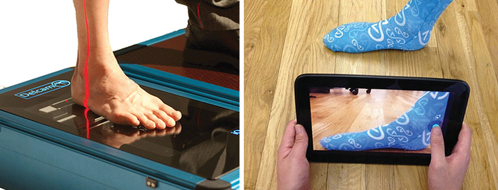
Robinson, who also suffers from chronic back pain caused by spinal stenosis, said that although he may not be able to swing a racquet anymore, he still can still fix things and do projects around the house.

“With my lack of dexterity, it takes me three times as long, and I have to use both hands to pick things up. I have to be more patient and inventive at times,” he said. “But my attitude on life is to be positive.”

Robyn Parets is a freelance writer in the Boston area.

Photos by Susan Ruediger.

The argument for library-based orthotic prescription

$
0
0

6Delcam-Sidebar-Picture1-copyBy Jordana Bieze Foster

Much of the discussion at the 2015 Ortho Technology Forum (OTF) focused on various ways in which the future of the orthotic industry will require adapting to new technologies. But the event’s final presenter challenged OTF attendees to consider the possibility that the future of the industry will also require reinventing the entire process of orthotic prescription.

Scott Marshall, co-owner of KLM Laboratories in Valencia, CA, noted that industry professionals who have been making custom foot orthoses have likely noticed, as he has, that a majority of the devices fall into recognizable and predictable shape categories. This phenomenon makes the industry particularly well-suited, he said, for a scientific library system in which most devices are made from premanufactured shells or positives or pre-engineered digital shape files.

“What if certain shapes are used over and over again? This is what optometrists do; that’s why you can get glasses in about an hour,” Marshall said.

What makes the proposed library system scientific is that the shape categories would be defined by specific measurements calculated from years of data on devices that were created using more traditional means of orthotic design and manufacturing.
KLM’s ongoing analysis of its database of 367,755 plaster casts and digital foot scans has revealed four key measurements that dictate device shape:

  1. Length from the bisection of the first metatarsal head to the most proximal aspect of the heel;
  2. Apex of medial arch height along the bisection of the first metatarsal shaft when the cast is placed in the balanced or prescribed position;
  3. Apex of lateral arch height when the cast is placed in the balanced or prescribed position; and
  4. Heel width across the middle of the heel, at the widest part.

After identifying the range for each measurement that accounted for the majority of devices in the database, Marshall and colleagues estimate that a library system of 4680 reproducible foot shapes would accommodate most patients’ needs. The library models could also easily be adjusted, mathematically and objectively, by increasing or decreasing one of the four key measurements.

This type of system could allow most patients to get high-quality orthotic devices at a single visit, and at a lower cost than with current processes, Marshall said. It could also provide a scientific means of tracking and studying the performance of different orthotic configurations on the same patient or on patients with particular deformities.

“The time has come to revisit how we prescribe orthotic devices, and to redefine what a custom orthotic is,” Marshall said.

The future of footwear and orthoses is here. Now what?

$
0
0

By Jordana Bieze Foster

A shift in tone was apparent at this year’s Ortho Technology Forum (OTF), and not just because the focus of the event has been expanded to include design and manufacturing technologies for footwear as well as foot orthoses. Speakers and attendees are no longer just speculating about how technology will change the footwear and foot orthosis industries—because those changes are already occurring in mainstream, high-profile ways.

The key difference between 3D laser scanning (left) and photogrammetry (right) is accuracy, experts say.

The key difference between 3D laser scanning (left) and photogrammetry (right) is accuracy, experts say.

The questions now focus on how foot care specialists’ role will change in an industry in which start-up companies often champion technical bells and whistles at the expense of accuracy and clinical relevance, and how clinicians themselves can use technology to their advantage to stay competitive.

“The war has started,” said Chris Lawrie, Healthcare Business Development Manager for Birmingham, UK-based Delcam Healthcare Solutions, which organized the April event held in Vancouver, Canada.

Lawrie was speaking specifically about how the growing accessibility of 3D printing technology has created a market for entrepreneurial footwear and orthoses manufacturers who are less con­- cerned with clinical effectiveness than their bottom line. But the assessment could as easily be applied to other aspects of technology being used by entrepreneurs to battle clinicians for a share of those markets.

“There isn’t a part of our industry that isn’t being affected by technology right now,” said Graham Archer, CPed(C), vice president of pedorthic services at Kintec Footlabs and president of Kiwi Software Solutions, both in Vancouver, in an OTF presentation.

The challenge for clinicians, and for other players in the footwear and orthotics markets for whom quality is a priority, will be to prove that new technologies aren’t just for newcomers. OTF presenters detailed multiple ways in which new tools and processes can help even established labs and clinics become more efficient, more accurate, and more profitable.

“The key is how to get around the paradigm shift to take advantage of things like new materials and new design opportunities,” Lawrie said.

3D printing

Feetz prototype custom shoes

Feetz prototype custom shoes

It’s tempting to dismiss 3D printing, also known as additive manufacturing, as the province of start-up companies looking to make a quick buck off drugstore-grade insoles or creative types designing futuristic-looking shoes that nobody in the real world would ever wear. But 3D printing is also being employed by a number of companies in ways that could potentially have much more practical and even clinical applications.

Chattanooga, TN-based Feetz is currently in the beta-testing phase of its personalized shoe business, which aims to 3D-print individual pairs of shoes based on either an in-store scan or on customers’ digital photos of their feet taken with the company’s app.

“One in three consumers are seeking personalization in their shoe purchases, and one in five Americans have foot issues that affect their shoe purchases,” Feetz CTO Nigel P. Beard said in an OTF presentation, while modeling a bright green prototype pair of Feetz shoes. “Our motto is: ‘You’ll never try on another pair of shoes again.’”

With few footwear-specific materials available that are compatible with 3D printing, the company ended up developing its own materials, ranging from ceramics to antimicrobials to scented polymers, Beard said.

Future directions, he said, include scanning shoe wear patterns and using 3D printing to build up material in the new pair to offset the expected wear, shoes that provide gradual orthotic correction along the lines of Invisalign orthodontic correction, and 3D-printing electronic circuits into footwear to provide feedback on how the shoe is affecting gait.

“I’m hoping we can not only change the face of regular footwear but also give you [OTF attendees] some different things to offer your patients,” Beard said.

Delcam has partnered with Eden Prairie, MN-based 3D printing company Stratasys to develop hybrid shoes with 3D-printed soles and heels and a leather upper, created using Delcam’s footwear design software. Prototype shoes made using this process were exhibited at SIMAC, an international exhibition of machines and technologies for the footwear and leathergoods industries, held in February in Milan, Italy. A similar process can also be used to create a conventional shoe with a 3D-printed insole, Lawrie said.

This type of evolution in footwear design and manufacturing may ultimately lead to a “lastless shoe,” he said.

Some technologies in which a tablet-based scanning device is moved around the foot, such as those utilizing the 3D Occipital Structure Sensor (sample image at right), approach the accuracy of more conventional laser scanners.

Some technologies in which a tablet-based scanning device is moved around the foot, such as those utilizing the 3D Occipital Structure Sensor (sample image at right), approach the accuracy of more conventional laser scanners.

“If we can create a digital last and three-D print the whole shoe, we’ll never need an actual last. That’s where the real savings are going to come from,” Lawrie said.

Other OTF presenters encouraged attendees to think about the possibilities of integrating 3D printing with other technologies to improve the manufacturing process. Robotics, for example, can improve efficiency and free up staff members to perform more complex tasks, and has the potential to help take 3D printing to the next level, according to Tom Bentley, owner of Waukesha, WI-based Robotic Solutions.

Potential uses for robotic automation in orthotic manufacturing include milling, gluing, assembly, sanding, packaging, and shipping, Bentley said.

“When you add robots to the equation for a milling function, even if it’s just to do that one function, you may be more cost effective. But what you want to do is look beyond the milling function at the four or five steps surrounding it. That’s how the robot really makes a difference,” he said.

Combining robotic technology and 3D printing is a logical next step, Bentley said.

“You could have a robot working with a 3D printer—loading it, for example,” he said. “That’s not a future thing. That’s something you can do now.”

Scanning

Just as entrepreneurial companies are jumping on the 3D printing bandwagon, they are also helping to popularize smartphone- and tablet-based scanning technologies for the design of what’s often billed as “custom” footwear and orthotic devices.

Clinicians and orthotic lab managers have years of experience sifting through the range of foot scanning devices on the market and determining the extent to which resolution, accuracy, and the ability to scan the entire foot justify the additional expense of high-end scanners. But now they’re faced with the prospect of competing with businesses that in some cases are creating footwear devices from 2D “scans” taken by the consumers themselves.

“As footcare professionals, we look at this and wonder how they’re going to make a true orthotic using this methodology. But this is out there, and these companies are going straight to the consumer and claiming they’re providing custom orthotics,” Archer said. “We need to be on top of this as an industry, and be able to answer questions from our patients and from insurance companies.”

The most critical issue, OTF presenters agreed, is accuracy.

“Don’t underestimate the role accuracy plays in what you’re doing when it comes to scanning technology,” Lawrie said.

Some of the technologies in which a mobile scanning device is moved around the foot, such as those utilizing the 3D Occipital Structure Sensor, approach the accuracy of more conventional laser scanners that require placing the foot against a flat scanner surface. But photogrammetry-based mobile scanning systems, in which a software algorithm translates 2D photographs of the foot into a 3D image, are unlikely to produce an accurate representation of foot structure, Archer said.

“There’s error throughout the entire process,” he said.

And at least one of the new foot-focused start-ups has made scanning accuracy a priority. At the Westfield North County Mall in Escondido, CA, customers can visit the Yooshu retail kiosk to have custom flip-flops made while they do the rest of their shopping. Each pair is designed based on a 3D laser scan (Littleton, CO-based Yooshu has been partnering with Delcam for the last year) and milled by a robot, CEO Scott Goldie said in an OTF presentation.

The result is a flip-flop with a customized sole, which can be further personalized with artwork. The products sell for $29 to $41 per pair, depending on materials; artwork is another $5. At the time of the OTF event, the first Yooshu location had been open for business for three weeks, and was already seeing repeat customers and referrals, Goldie said.

The company invested in laser scanning technology specifically to get an accurate scan of each patient’s toes, he said, but there are plans to also use the scan data to position dorsal straps and to save customer scans to facilitate easy ordering of duplicate pairs.

“The scanner has more capability than we’re using right now,” Goldie said. “The robot is expensive, but it’s getting cheaper and cheaper. Our real investment has been in software and training.”

Another future direction includes offering a more sophisticated product for customers who wear foot orthoses.

“Of the customers who decline to purchase after the pitch, fifteen to twenty-five percent are looking for orthotic features we don’t offer yet,” Goldie said. “We’re not interested in going ‘full medical,’ but it’s not far for us to go from our flip-flop to a truly orthotic flip-flop.”


Childhood obesity and OA: Can early care reduce risk?

$
0
0

8peds-Obesity-iStock9809464-copyBy Erin Boutwell

Osteoarthritis (OA) risk factors and symptoms seen in adults have been found in obese kids, who often have musculoskeletal pain. Weight loss may help, but preventing OA may also require gait and exercise interventions, particularly those that reduce pain that leads to inactivity.

Osteoarthritis is an extremely common musculoskeletal disorder. In 2008, an estimated 27 million US adults had clinical OA in one or more joints.1 OA may be caused by mechanical, systemic, and genetic factors.2 It represents the failure of a joint caused by abnormal mechanical loads, and the ability to comprehend this disorder requires a biomechanical perspective.3

No cure exists for OA,4 though clinicians often emphasize weight loss and exercise because of the clear association between musculoskeletal issues and obesity in adults.5-7 However, the applicability of adult-centered research findings to the treatment of obese children—who do not yet have OA but appear to be at increased risk—is problematic.

It may be possible to reverse some musculo- skeletal symptoms seen in obese children, particularly those associated with OA in adults: pain, cartilage damage, and gait deviations

Simone Gill, PhD, OTR/L, assistant professor in the Department of Occupational Therapy and director of the Motor Development Laboratory at Boston University, outlined a major issue in extending adult OA research findings to a pediatric population. “Children are still growing and developing, and we know very little about how obesity impacts changes in their development,” she said.

Because the rising worldwide population of persons classified as “overweight” (body mass index [BMI] ≥ 25 kg/m2 and < 30 kg/m2) or “obese” (BMI ≥ 30 kg/m2) is expected to contribute to increased OA incidence,4 it’s important to consider early intervention in heavy children and adolescents. Assessment of body fat in children is not calculated using an absolute BMI, as in adults, but rather using age- and gender-specific percentiles; in children aged 5 to 19 years, “overweight” typically corresponds to a BMI between the 85th and 94th percentile, and “obese” is typically associated with a BMI greater than the 95th percentile.8

No data have established a direct link between OA and obesity in children, but researchers are beginning to uncover evidence of musculoskeletal complaints and injuries that are precursors to adult OA in the younger obese population.

Obesity in children

Currently, the connection between OA later in life and childhood obesity is primarily hypothetical. Marienke van Middelkoop, PhD, assistant professor at the Erasmus MC Medical University Department of General Practice in Rotterdam in the Netherlands, offered an explanation of the mechanism by which obesity could be a risk factor for development of musculoskeletal issues in children.

“Obesity could indeed be a cause of increased strain on joints during weight bearing,” she said. “The [same] mechanism has also been suggested for the association between obesity and osteoarthritis of weight-bearing joints such as the hip and the knee.”

Research has linked knee OA to excessive forces transmitted across a joint because of high body weight, potentially leading to cartilage breakdown and progressing to OA.9 Loading alone, however, doesn’t explain why hand OA is also more prevalent in obese individuals.9

“There are also other potential disease pathways suggested [for obesity-related osteoarthritis], including inflammation and hormone secretion,” van Middelkoop said.

Musculoskeletal pain

Children, in general, are at a high risk for musculoskeletal pain; studies have reported that between 18% and 32% of children experience pain at least once a week.10-12 The exact mechanism behind this pain is unknown, but it may be related to overuse.13 A systematic review coauthored by van Middelkoop suggests the occurrence of musculoskeletal pain may be even higher in heavier children than in the general population.5 In the review, van Middelkoop found the overall prevalence of musculoskeletal pain was 26% higher in overweight children than in normal-weight children, and that low back pain in particular was 42% more common in overweight children.

Cartilage lesions

Perhaps the strongest link between obesity and musculoskeletal degeneration comes from a series of studies using magnetic resonance imaging (MRI) to examine the knee joints of morbidly obese children and adolescents.14,15 (Morbid obesity was defined as BMIs exceeding the 99.5th percentile for participants’ age and gender.) These studies, coauthored by Harald Widhalm, MD, of the Department of Trauma Surgery at the Medical University of Vienna in Austria, demonstrate the possibility that children aged as young as 9 years may show symptoms of joint degeneration. In a 2012 study, Widhalm and colleagues found that 100% of 20 morbidly obese children aged 9 to 19 years had cartilage lesions in at least one knee compartment.

“These [morbidly obese] patients often present with severe deviations of the leg, showing cartilage pathologies…in the medial joint,” said Widhalm, who went on to warn, “The fact that these patients are often developing cartilage lesions even at that young age is a big problem. We also do not know…if cartilage lesions in these patient types are reversible.”

Articular cartilage lesions have been related to long-term joint issues such as adult OA.16 While a direct causative link between these lesions and obesity has not been established, Widhalm et al reported their prevalence seems to correlate with musculoskeletal pain symptoms commonly found in obese children.17

Knee malalignment

Poor frontal-plane knee alignment can play a large role in joint loading. In a 2014 Rheumatology study van Middelkoop and others noted a significant increase in the risk of developing radiographic knee OA in middle-aged overweight women with knees in varus alignment (odds ratio of 3.3), finding a more modest association in women with valgus knee alignment (odds ratio of 2.8).18

Sarah Shultz, PhD, ATC, a lecturer in exercise and sport science at Massey University in Wellington, New Zealand, noted, “If the child is choosing to be in genu valgum in order to relieve some pressure medially…then they are inadvertently placing themselves at greater risk for osteoarthritis to the lateral aspect of the knee.”

Nor is the knee the only critical joint to consider in obese children. “The foot and ankle [complex] is the first line of defense against the extra ground reaction forces; combined with the architecture of the foot [small bones, ligaments, arches], it makes the foot highly susceptible to changes due to mass,” she said.

Another potential issue in obese children is that their increased body mass may result in gait compensations that exacerbate these lower limb malalignments and may increase the risk of OA.19 Gill elaborated, “Obese children tend to walk more slowly, and then try to stabilize their bodies…by keeping their feet on the ground for longer periods of time during the gait cycle.” Previous work documents that obese children may walk at slower speeds,20 increase their step widths,20 and experience higher joint powers at the hip and knee during loading response.21

Treatment

Weight loss. Weight loss is commonly recommended in adults as a method of reducing abnormal loading on lower limb joints,9 and many doctors and scientists advocate multidisciplinary weight-loss approaches. Widhalm coordinates such weight reduction programs. “It is very important to have a multiprofessional team; otherwise, there is no chance to help these patients,” he said.

However, is weight loss alone enough?

Sharon Bout-Tabaku, MD, assistant professor of pediatrics at Nationwide Children’s Hospital in Columbus, OH, said, “The question is: Can you can make people’s outcomes even better in regards to pain and risk of developing osteoarthritis, if—along with weight loss—you can strengthen their muscles or if you can help train them to walk better?”

Exercise & physical therapy. Bout-Tabaku gave an example of a weight-loss concurrent therapy, suggesting, “Obese kids have a lot of truncal mass, and it’s possible that truncal mass, even though it improves with weight loss, still causes some instability. Thus, strengthening of the core and lower extremity muscle groups may help the downstream effects of injury to the knee.”

Gill also suggested that gait pattern deviations could be addressed through exercise interventions. “Changes in muscle quality can happen with strength training,” she said, then highlighted the importance of cardiovascular fitness. “Another possible benefit in terms of aerobic activity in particular is that it not only helps burn fat, but also contributes to increased blood flow in the brain, which might be related to some of the issues of motor planning in that population.”

Shultz observed, “I think it’s important to include physical activity in any weight-management program, but physical limitations should be considered. I am a big believer in resistance training for obese youth and think that activities like boxing or aquatics are great alternatives to the more conventional activities [such as running].” Alternative activities are important to consider in obese children because the higher mechanical joint powers [the rate of mechanical work performed at the joint] they experience during exercises such as running may reduce their motivation for regular participation.21

Orthotic devices. Studies have evaluated various orthotic interventions for reducing pain and redistributing load within the knee joint in adults with OA, but the efficacy of such devices in adult knees is inconclusive at best. Several studies have investigated the effects of lateral wedges on peak knee external adduction moment, a measure often used to approximate knee loading,22 and have reported peak moment reductions of 5.8%23 and 8.7%24 with a laterally wedged insole compared with a neutral insole during walking in subjects with medial knee OA. The effects of lateral wedges on pain, however, have been inconsistent.25,26 Also, there are few high-quality studies that have evaluated the use of knee bracing in patients with knee OA;27 however, some investigations have reported improved walking distance28,29 and pain relief28-30 with a knee brace compared with no brace.

The potential influence of orthotic devices is an as yet unexplored avenue of research in correcting joint malalignment or preventing OA risk factors in children in adolescents. “Both knee braces and wedged insoles reduce the medial knee joint load [in adults], but the preventive effects have never been studied,” noted van Middelkoop. “It is so far unknown if you can prevent osteoarthritis with these interventions in high-risk groups.”

Reversibility. The ability to reverse obesity-related disease states that occur in children and adolescents is an important topic to explore. Bout-Tabaku said drawing a distinction between musculoskeletal complaints and OA in children is key. She queried, “What does ‘disease’ mean? Does ‘disease’ mean just having pain? Or does ‘disease’ mean having osteoarthritis?”

She added, “We don’t have any data saying that kids already have osteoarthritis.” All that has been reported, she said, is the existence of “some lesions in obese children that may be similar to what people see in adults who have very early stage osteoarthritis,” a reference to Widhalm’s previously cited detection of articular cartilage lesions in morbidly obese children.14,15

Therefore, “reversing” OA in children may not be a meaningful goal. It may be possible, however, to reverse some of the musculoskeletal symptoms demonstrated by obese children, particularly those that have been associated with OA in adults: pain, cartilage damage, and gait deviations. But how reversible are these symptoms?

Investigators like Gill are optimistic about the ability of weight loss and exercise regimens to reverse some of the musculoskeletal disease and damage reported in obese children and adolescents, including poor muscle quality and gait abnormalities. Improvements in gait (eg, increased step lengths, increased walking speeds, and reduced step width) have been reported in adults who have undergone bariatric surgery in as little as three months postsurgery.31

Regarding the reversal of cartilage damage, though, Gill was not as optimistic. “If you decrease the load that is being applied to those joints, then you might be able to minimize the wear and tear that we all experience in terms of lost cartilage over time. Because obesity exacerbates that,” she said. “That [cartilage loss] might not be reversible, per se, but you can slow how much that is happening.”

Gill’s thoughts build on the findings of a seminal 2005 Arthritis & Rheumatism study in which the authors found that in adults, for each pound of weight loss, four fewer pounds of force are applied to the knee joint.32 However, a direct causative link between reduction in knee load and slowed disease progression has yet to be established.

Nor is it clear exactly where the threshold between reversible and permanent joint damage lies. Bout-Tabaku asked, “When you’re done growing, does obesity confer greater or less of a risk than when you’re still growing?”

The idea that a child’s stage of development could affect obesity-related risk factors is a finding of Bout-Tabaku’s 2015 Journal of Rheumatology paper, in which she reported that pubertal stage (measured by Tanner stage) influenced knee alignment in obese children.33 In particular, knee alignment was comparable between obese and normal-weight children at Tanner stages 2 and 3, but, at Tanner stages 4 and 5, she found a greater valgus alignment in obese compared with normal-weight children (See “Obese children develop knee malalignment as they mature,” LER: Pediatrics, May 2015, page 5).

Bout-Tabaku emphasized the importance of pain reduction in obese kids, saying, “Pain itself is a disease state. We know that obese kids have more prevalent pain than nonobese kids.” She found that 76% of obese adolescents participating in the Teen–Longitudinal Assessment of Bariatric Surgery (Teen-LABS) study had pain at baseline.34

The next, ongoing phase of the Teen-LABS study involves investigating whether weight loss can reduce the pain incidence in these children. Bout-Tabaku said, “If kids are having pain, they then go into this vicious cycle of being more sedentary and deconditioned, putting them at risk for falls, abnormal joint mechanics, and injury to their joints.” In adults, weight loss has been associated with a reduction in joint loading,35 inflammation,35,36 and pain.31,35,36

The big picture

A common theme among all researchers and clinicians in this area was the information gap with regard to OA risks in this young population. Even more mysterious is how to identify root causes versus downstream effects.

Gill is convinced longitudinal studies are needed to parse some of the cause-and-effect relationships proposed between obesity and poor motor function over time. Bout-Tabaku added that a longitudinal study tracking obese and nonobese individuals over time who start with a normal knee alignment could be extremely informative.

These numerous unknowns, combined with the detrimental effects of OA, make it clear that intervening early, before musculoskeletal problems develop, is key. As Widhalm puts it, “For sure, prevention of morbid obesity is the best strategy.”

Erin Boutwell is a freelance writer based in Chapel Hill, NC.

REFERENCES

1. Lawrence RC, Felson DT, Helmick CG, et al. Estimates of the prevalence of arthritis and other rheumatic conditions in the United States. Part II. Arthritis Rheum 2008;58(1):26-35.

2. Bout-Tabaku S, Best TM. The adolescent knee and risk for osteoarthritis – an opportunity or responsibility for sport medicine physicians? Curr Sports Med Rep 2010;9(6):329-331.

3. Dieppe P. Developments in osteoarthritis. Rheumatology 2011;50(2):245-247.

4. Fransen M, McConnell S, Harmer AR, et al. Exercise for osteoarthritis of the knee. Cochrane Database Syst Rev 2015;1:CD004376.

5. Paulis WD, Silva S, Koes BW, van Middelkoop M. Overweight and obesity are associated with musculoskeletal complaints as early as childhood: a systematic review. Obes Rev 2014;15(1):52-67.

6. Kortt M, Baldry J. The association between musculoskeletal disorders and obesity. Aust Health Rev 2002;25(6):207-214.

7. Butterworth PA, Landorf KB, Smith SE, Menz HB. The association between body mass index and musculoskeletal foot disorders: a systematic review. Obes Rev 2012;13(7):630-642.

8. Barlow SE, Expert Committee. Expert committee recommendations regarding the prevention, assessment, and treatment of child and adolescent overweight and obesity: summary report. Pediatrics 2007;120 Suppl 4:S164-192.9. Felson DT. Does excess weight cause osteoarthritis and, if so, why? BMJ 1996;55(9):668-670.

10. Mikkelsson M, Salminen JJ, Kautiainen H. Joint hypermobility is not a contributing factor to musculoskeletal pain in pre-adolescents. J Rheumatol 1996;23(11):1963-1967.

11. Leone V, Tornese G, Zerial M, et al. Joint hypermobility and its relationship to musculoskeletal pain in schoolchildren: a cross-sectional study. Arch Dis Child 2009;94(8):627-632.

12. Smith SM, Sumar B, Dixon KA. Musculoskeletal pain in overweight and obese children. Int J Obes 2014;38(1):11-15.

13. Kaspiris A, Zafiropoulou C. Growing pains in children: epidemiological analysis in a Mediterranean population. Joint Bone Spine 2009;76(5):486-490.

14. Widhalm HK, Marlovits S, Welsch GH, et al. Obesity-related juvenile form of cartilage lesions: a new affliction in the knees of morbidly obese children and adolescents. Eur Radiol 2012;22(3):672-681.

15. Widhalm HK, Seemann R, Hamboeck M, et al. Osteoarthritis in morbidly obese children and adolescents, an age-matched controlled study. Knee Surg Sports Traumatol Arthrosc 2014 May 20. [Epub ahead of print]

16. D’Anchise R, Manta N, Prospero E, et al. Autologous implantation of chondrocytes on a solid collagen scaffold: clinical and histological outcome after two years of follow-up. J Orthopaed Traumatol 2005; 6(1):36-43.

17. Mohseni-Bandpei MA, Bagheri-Nesami M, Shayesteh-Azar M. Nonspecific low back pain in 5000 Iranian school-age children. J Pediatr Orthop 2007;27(2):126-129.

18. Runhaar J, van Middelkoop M, Reijman M, et al. Malalignment: a possible target for prevention of incident knee osteoarthritis in overweight and obese women. Rheumatology 2014;53(9):1618-1624.

19. Shultz SP, D’Hondt E, Fink PW, Lenoir M, Hills AP. The effects of pediatric obesity on dynamic joint malalignment during gait. Clin Biomech (Bristol, Avon) 2014;29(7):835-838.20. Deforche BI, Hills AP, Worringham CJ, et al. Balance and postural skills in normal-weight and overweight prepubertal boys. Int J Pediatr Obes 2009;4(3):175-182.

21. Shultz SP, Hills AP, Sitler MR, Hillstrom HJ. Body size and walking cadence affect lower extremity joint power in children’s gait. Gait Posture 2010;32(2):248-252.

22. Barrios JA, Butler RJ, Crenshaw JR, et al. Mechanical effectiveness of lateral foot wedging in medial knee osteoarthritis after 1 year of wear. J Orthop Res 2013;31(5):659-664.

23. Chapman GJ, Parkes MJ, Forsythe L, et al. Ankle motion influences the external knee adduction moment and may predict who will respond to lateral wedge insoles?: an ancillary analysis from the SILK trial. Osteoarthritis Cartilage 2015;23(8):1316-1322.

24. Butler RJ, Marchesi S, Royer T, Davis IS. The effect of a subject-specific amount of lateral wedge on knee mechanics in patients with medial knee osteoarthritis. J Orthop Res 2007;25(9):1121-1127.

25. Erhart JC, Mündermann A, Elspas B, et al. Changes in knee adduction moment, pain, and functionality with a variable-stiffness walking shoe after 6 months. J Orthop Res 2010;28(7):873-879.

26. Maillefert JF, Hudry C, Baron G, et al. Laterally elevated wedged insoles in the treatment of medial knee osteoarthritis: a prospective randomized controlled study. Osteoarthritis Cartilage 2001;9(8):738-745.

27. Duivenvoorden T, Brouwer RW, van Raaij TM, et al. Braces and orthoses for treating osteoarthritis of the knee. Cochrane Database Syst Rev 2015;3:CD004020.

28. Sattari S, Ashraf AR. Comparison the effect of 3 point valgus stress knee support and lateral wedge insoles in medial compartment knee osteoarthritis. Iran Red Crescent Med J 2011;13(9):624-628.

29. Brouwer RW, van Raaij TM, Verhaar JA, Coene LN, Bierma-Zeinstra SM. Brace treatment for osteoarthritis of the knee: a prospective randomized multi-centre trial. Osteoarthritis Cartilage 2006;14(8):777-783.

30. Kirkley A, Webster-Bogaert S, Litchfield R, et al. The effect of bracing on varus gonarthrosis. J Bone Joint Surg Am 1999;81(4):539-548.

31. Vincent HK, Ben-David K, Conrad BP, et al. Rapid changes in gait, musculoskeletal pain, and quality of life after bariatric surgery. Surg Obes Relat Dis 2012;8(3):346-354.

32. Messier SP, Gutekunst DJ, Davis C, DeVita P. Weight loss reduces knee-joint loads in overweight and obese adults with knee osteoarthritis. Arthritis Rheum 2005;52(7):2026-2032.

33. Bout-Tabaku S, Shults J, Zemel BS, et al. Obesity is associated with greater valgus knee alignment in pubertal children, and higher body mass index is associated with greater variability in knee alignment in girls. J Rheumatol 2015;42(1):126-133.

34. Bout-Tabaku S, Michalsky MP, Jenkins TM, et al. Musculoskeletal pain, self-reported physical function, and quality of life in the Teen-Longitudinal Assessment of Bariatric Surgery (Teen-LABS) Cohort. JAMA Pediatr 2015;169(6):552-559.

35. Messier SP, Mihalko SL, Legault C, et al. Effects of intensive diet and exercise on knee joint loads, inflammation, and clinical outcomes among overweight and obese adults with knee osteoarthritis: the IDEA randomized clinical trial. JAMA 2013;310(12):1263-1273.

36. Richette P, Poitou C, Garnero P, et al. Benefits of massive weight loss on symptoms, systematic inflammation and cartilage turnover in obese patients with knee osteoarthritis. Ann Rheum Dis 2011;70(1):139-144.

Growing pains: Adapting O&P devices to maturing patients

$
0
0
Photo courtesy of Cascade Dafo

Photo courtesy of Cascade Dafo

By Shalmali Pal

Accommodating growth without compromising fit and function is a challenge for practitioners who prescribe orthotic devices for young patients. Adjustments and add-ons—as well as educating parents about expected changes—can make for smoother transitions.

YouTube is a treasure trove of feel-good videos featuring children walking for the first time with the help of O&P devices. There’s 2-year-old Kayden, 1-year-old Samantha, and 14-year-old Zoe, who lost her left leg below the knee in a jet skiing accident.

But these inspiring images capture only one moment in each child’s journey. What happens down the road as Kayden, Samantha, and Zoe mature and outgrow their devices?

The need to replace an O&P device on a regular basis has its challenges for practitioners, patients, and parents. The patient who has just become accustomed to a device has to start all over. Parents want the best for their children but know these replacements can come with a hefty price tag. And that means the practitioner must attempt to optimize fit and function for the moment, make adjustments over time if possible, and, ultimately, determine when a completely new device is necessary.

LER: Pediatrics spoke with O&P experts on how to handle these issues, including how to accommodate growth without compromising fit and function.

While not a technical issue, patient and parent education is key to ensuring that everyone understands the purpose of a device and its projected longevity.

“We are always considering growth: How long can they wear it? What can we do to try to make it last longer?” said Leigh Davis, CPO, LPO, of Children’s Healthcare of Atlanta, and a board member of the American Academy of Orthotists and Prosthetists. “At the same time, since growth is so variable, you want to plan for what you see now rather than what might happen. You want to plan for care today versus planning for something that might happen in the future.”

Growth spurts

Of course, all children grow, but as Davis pointed out, not all kids grow at the same rate, even during infant, toddler, and adolescent years, which are known for growth. And children who require O&P devices often have compromised skeletal systems and musculature because of their underlying diagnosis, and that may affect their growth schedule.

Tony Wickman, CTPO, chief executive officer of Freedom Fabrication in Havana, FL, noted that bones tend to grow faster linearly than they do circumferentially. Unlike in adults, in whom changes tend to be more global, changes in kids may happen in a piecemeal fashion. For instance, a child may experience linear growth of the feet rather than medial-lateral growth, but without any major growth in the rest of the lower limbs.

Photo courtesy of Allard USA

Photo courtesy of Allard USA

Davis agreed that linear growth is more of a concern in kids who may not build muscle mass as quickly as typically developing children.

“So you don’t often see a really large increase in calf circumference in kids with certain disabilities,” she explained. “But let’s say their ankle-malleolar width increases so much that it’s painful to put the brace on or they can’t even put it on at all. They’d have to come in so that we could make adjustments to the device.”

At the O&P service at Ann & Robert H. Lurie Children’s Hospital of Chicago, Nikta Pirouz, CPO, and colleagues make ankle foot orthoses (AFOs) for children as young as a day old.

“Any presentation that requires an orthosis at that young an age requires a good fit even though these children aren’t ambulatory,” she said.

Many children who require O&P devices have underlying neurological conditions that can impact their ability to understand the purpose of the devices, use them properly, and express when the device is causing discomfort, possibly because of a change in body habitus.

Pirouz noted that standard clinical growth charts don’t really apply to kids who require devices, even if they don’t have underlying conditions.

“Even for typical kids, those charts are an aggregate. The chart will tell me where the child will be in a year, but it’s not going to tell me exactly when that will happen for each individual child. A week from now? Over the summer? That’s why follow-up is so important,” she said.

Finally, just as in the general population, childhood obesity is a problem in O&P patients. Pirouz pointed out that weight gain will most likely necessitate adjustments so that a device doesn’t pinch or dig into “redundant soft tissue.” But those adjustments don’t have to be complex.

“Whether it’s weight gain, or volume change that is part of the disease presentation, you can address that,” she said. “You can add a tongue to contain tissue. You can flare. You can pad. You can try interfaces to contain the tissue better.”

Don’t fit for the future

It’s not worth trying to fit a device for potential growth because it will compromise fit in the here and now, experts agreed.

“I want the device as fitted as much as possible to the child’s current shape and size,” Wickman emphasized. “What I don’t like to see is a big, sloppy fit in hopes that the child will grow into it. Then you lose control of the fit and possibly compromise the device’s
efficacy.”

8PEDSgrowin-AllardShoot4.14.13-1773

Photo courtesy of Allard USA

Davis concurred. For example, she said she wouldn’t typically build extra padding into an AFO that could be removed at a later time, because that can add bulk and compromise fit. She would leave the footplate a centimeter longer than the toes to accommodate for growth—but no longer, as that can cause problems with tripping or may require a shoe that’s a size too big. Footplate size can be misleading to parents as well, she said.

“A lot of parents will rely on the footplate as a measure of growth. So if they wait [to replace the device] until the toes are hanging all the way over the footplate, then the height of the AFO becomes a concern,” she said.

Pirouz agreed that achieving maximum fit and function in the present is paramount. Adjustments for growth can be made, but in the right context.

“For instance, with a night AFO, the patient is not ambulating, so you can make that footplate as long as you want in order to accommodate for growth,” she said. “But if you think about the height of the device, that can only be so tall, otherwise it may make the brace uncomfortable.”

Adjustments, add-ons, education

Adding that extra centimeter to a footplate is just one of many tactics practitioners use to maintain a quality fit while also accommodating for growth. But no single approach will work well for all children.

“You really have to look at the individual child and their diagnosis, their size, and their level of tolerance,” Davis said. “Is the weight of the device an issue for them? Is cosmesis an issue? Those sorts of things have to be taken into consideration.”

Wickman also cited diagnosis as a driving factor.

“With a diagnosis that’s relatively easy to manage, like genu valgum at the knee, that takes relatively gross pressures to manage and can be done with a minimum of contact,” he explained. “So in circumstances like that, it’s quite easy to manage for growth. The less surface that you have touching the patient, the more room you have for growth.”

But treating a condition like clubfoot is another matter, because the volume and circumference of the orthoses have to be managed in addition to the length, he added.

LER asked the O&P specialists specifically about a number of common techniques.

Leaving room to grow. Again, no one advocated a compromised fit for future growth. As Davis mentioned, she may leave a bit of room on an AFO footplate, but not at the top of the brace as it would hinder the child’s ability to bend their legs. Building extra width or circumference into a device also may compromise that device’s ability to control movement.

“Most of the kids are going to have pretty serious frontal plane involvement, so you need to be able to control supramalleolarly,” Davis said.

Infants grow very quickly so leaving room to grow can be tricky, Wickman said.

“There’s an old adage in O&P about growth adjustment: ‘The more you need, the less you can have.’ Very small children are going to need a lot of growth adjustment, but if they are smaller, then we can’t build in a lot for growth because achieving the best fit doesn’t give us a lot of room to work with,” he said.

Pirouz noted that pediatric devices generally don’t encapsulate the entire limb so allowances for growth can be made. This may mean using different interfaces to gain the best fit or using different tensions on the straps.

Adding toe pads or removable liners. Davis and Pirouz said they did not use removable liners because of the bulk they add, but have used toe pads to increase the length of a device.

Wickman acknowledged that removable liners can allow for extended device use in a growing child, but agreed that the added bulk and weight may not be desirable. Instead, he recommended using spot pads to cushion the bony prominences without adding bulk.

Heating/flaring to increase volume. Increasing volume by heating is a go-to protocol for making adjustments, Wickman said, and today’s thermoplastic materials can withstand heating if the circumference of the device needs to be increased. Another benefit of using spot pads rather than full liners is that the latter can get in the way of heating the device, he said.

Davis also supports heating to increase or decrease the circumference of a device, but she cautioned that doing so will also change the depth of the device in ways that could compromise its effectiveness.

“If I heat it out and it makes [the device] wider, it’s not as deep as it used to be,” she said.

Pirouz pointed out that it’s easier to maintain depth when decreasing the volume of a device than when increasing it.

“You can just cut the plastic away and maintain the depth,” she said. “If you are trying to increase the volume, then you will get a shallower device. As the calf portion of the AFO gets shallower from widening it [with heating], you can add a foam tongue to address that problem to match the volume of the device to the patient.”

Screwed-on joints vs internal or riveted joints. In general, Davis advised positioning screwed-on joints on the outside of the device so the practitioner has the option to move the proximal section superiorly.

With knee AFOs (KAFOs), she said she aims to build in the capability to bring the knee joint up for growth, whether that’s with Chicago screws or an attach plate.

“In either case, you can do growth adjustments, although the attach plate will add a lot of thickness,” Davis said. “We don’t do attach plates very often. More often, we move the holes and re-rivet or re-Chicago screw it.”

The use of growth adjustment bars will depend on the size of the child and how much weight he or she can tolerate, she added.

“If it’s a very small child, that may be more than they can handle,” Davis said. “Now, if it’s an adolescent, then a growth adjustment bar is probably a good idea. They have a lot of growth at that age, plus they are a bit bigger so that extra weight isn’t going to be that big a deal.”

In terms of adjustment bars, Wickman recommended stainless steel over aluminum as the former offers lighter weight but better durability, especially because active kids can be particularly rough on their devices. Stainless steel also will handle the stresses from repeated bending forces, for example in devices for genu varum or genu valgum, he said.

Pirouz said her group has not had any problems with internal joints, though she conceded that “you do have to extend your build-up to accommodate for that growth where the malleolae are going to move proximally.”

Prefab vs custom. The use of off-the-shelf (OTS), or prefabricated, devices may seem like an attractive option for growing children simply because they are less expensive than custom devices. But other factors also come into play.

Davis said her group does not use OTS devices very often in their pediatric population, in part because, in their experience, OTS devices aren’t always effective in kids who have triplanar deformities.

For Pirouz, whether a device is OTS or custom is less of an issue than its material construction.

“I think being able to make adjustments is the key, and some prefab devices allow you to do that while others don’t. It’s the same with some custom devices,” she explained. “Let’s say you have a custom carbon AFO that is laminated. Unless you build in padding that you can grind out, you really don’t have a lot of room to adjust that device.”

On the other hand, an OTS carbon AFO may have such a low profile, and such limited contact with the anatomy, that it can accommodate circumferential or linear growth, Pirouz said.

“Sometimes the referring specialist will just assume that a prefab device won’t work, and that’s not necessarily true,” she said.

Education. While it’s not a technical issue, patient and parent education is key to ensuring that everyone understands the purpose of a device and its projected longevity.

“A huge component of accommodating for growth is education—making sure the patient and the parents know what the device should look like when it’s properly fitted and when it may be time to come in for adjustments,” Pirouz stressed.

Pirouz said she and her colleagues start with very short follow-up periods of one to two weeks to establish compliance and a schedule for wearing the device. After that, follow-up appointments are scheduled every one to three months, depending on the child’s age.

“We always explain that we are shooting for the brace to last a year, but the priority is fit and function, so there is a chance that a new brace might be needed sooner,” she cautioned.

Davis makes sure to take measurements every time the patient comes for a follow-up visit so, should a new brace be necessary, she’ll have the data to back up that request.

“A lot of insurance companies will replace the device if there’s documented physiologic change,” she said. “So, let’s say the patient has gained ten pounds; that can be a valid justification to get a new brace.”

It also can be reassuring to parents who are concerned about the cost of replacing a device sooner rather than later.

On the other hand, practitioners may also find themselves contending with parents who will insist on a new device on a regular basis, especially if they believe that a newer device will work better and faster. Pirouz said that scenario is one she encounters more often than parents who are reluctant to get a new device. But if the desired clinical results are seen during regular follow-up, she said, then there may not be a need to jump immediately to a new brace.

Shalmali Pal is a freelance writer in Tucson, AZ.

Robotic gait training doesn’t wow young patients with CP

$
0
0
Photo courtesy of William Suarez/Holland Bloorview Kids Rehabilitation Hospital

Photo courtesy of William Suarez/Holland Bloorview Kids Rehabilitation Hospital

Kids, parents differ on ‘normal’ gait

By Brigid Galloway

Although physical therapists and parents often strive for attaining “normal” gait in children with neuromotor disorders, a new study from researchers at the University of Alberta in Edmonton, Canada, indicates that children undergoing robotic gait training therapy may not share the same enthusiasm for this goal.

The qualitative study assessed the expectations and experiences of five children aged 8 to 11 years with cerebral palsy (CP, gross motor function classification system levels II or III), who underwent robotic gait training with a treadmill-based gait trainer, and their parents. The trainer provides upright support while using robotic orthoses to move the patient’s legs. Its adjustable body-weight support allows children to use a more vertical, physiologically typical gait pattern.

Over about 10 weeks, the children underwent 16 sessions of robotic gait training, which involved stepping over blocks, active hip extension against resistance, and playing with a ball while walking. The sessions also included video games in which children had to do specific movements to advance in play.

The study underscores the need for research that focuses on the opinion and voices of young patients, who often get mixed messages.

Researchers interviewed children and parents after the child’s last robotic training session. They asked parents about their expectations, experiences, and perceived outcomes with regard to the robotic trainer and walking goals for their child. Questions for children were designed to get their perspective on walking, as well as how they felt about using the trainer.

Parents generally expressed interest in continuing robotic gait training, felt it made their children more confident, and assumed their children found the games engaging and that they valued being able to walk like a typically developing child.

The children had more mixed impressions of the robotic trainer, finding it alternately “fun,” “boring,” and even “uncomfor­table/painful.” Young respondents also did not echo their parents’ point of view on “normal” gait, said lead author Shanon Phelan, PhD, assistant professor of occupational therapy at the University of Alberta. “They often equated normal walking with their typical gait, weren’t always sure whether the robot was helping them, and didn’t universally find it fun or engaging.”

Parents thought robotic training, which took place at Holland Bloorview Kids Rehabilitation Hospital in Toronto, boosted children’s confidence, but the children didn’t express this, and some felt anxiety about engaging the technology. Also a prominent problem for children was skin irritation caused by the trainer’s straps.

Both children and parents, however, said they valued the interactions with their therapists regardless of the technology.

“This study calls for critical reflection on why and how one might engage children in gait-related rehabilitation in ways that bring children’s desires and expectations to the forefront,” said Phelan. “This challenges clinicians to rethink how gait-related interventions are presented to children and families and how goals and outcomes are framed. For example, if a child is ambivalent about the quality of his or her gait, or [already] sees his or her gait pattern as ‘normal,’ one might consider presenting the benefits of an intervention in ways that highlight what is meaningful to the child and reinforce their abilities and positive self-identity.”

The study underscores the need for research that focuses on the opinion and voices of young patients, who often get mixed messages, she said.

“People in their lives often reinforce the idea of social acceptance. They hear, ‘there is nothing wrong with the way you walk,’ yet they participate in therapy to work on walking more like their same-aged, nondisabled peers,” she said. “When we focus solely on the physical aspects of walking/gait training, we can forget about the social and emotional implications for children.”

Disability and Rehabilitation epublished the study on April 9.

Theresa Sukal Moulton, PT, DPT, assistant research professor at Northwestern University in Evanston, IL, has worked extensively with children with CP using therapeutic robotic devices. Despite the inability to generalize the feelings, perceptions, and goals of this small cohort of patients onto all children with gait disorders, the study brings significant considerations to light, she said.

“The mismatch between a parent’s and child’s goals and perceptions about the effectiveness of therapeutic modalities is not uncommon in my clinical experience,” said Moulton. “Pediatric clinicians have the unique challenge of treating the whole family, so the perspective of both parents and patients must be considered and balanced, in an age-appropriate way.”

Brigid Galloway is a freelance journalist in Birmingham, AL.

Source:

Phelan SK, Gibson BE, Wright FV. What is it like to walk with the help of a robot? Children’s perspectives on robotic gait training technology. Disabil Rehabil 2015 Apr 9:1-10. [Epub ahead of print]

Gait analysis for clubfoot may reveal long-term issues

$
0
0
Photo courtesy of MD Orthopaedics

Photo courtesy of MD Orthopaedics

Surgery more likely to alter gait

By Larry Hand

Children treated for idiopathic clubfoot by age 2 years may experience subtle changes in gait by the time they are aged 5 years, and nonoperative treatment may confer more normal movement than surgery, according to a recent study.

After treatment for idiopathic clubfoot at the Texas Scottish Rite Hospital for Children in Dallas, investigators analyzed data from 181 children seen for gait analysis when aged 2 and 5 years. Among 276 feet, 132 underwent Ponseti treatment, and 144 received French physical therapy (PT). By the time children were aged 5 years, 30 Ponseti and 61 PT feet had required surgery (47 feet before age 2 years, 44 between ages 2 and 5 years).

Among the surgically treated children, data collected at their second gait analysis showed only 17% of the Ponseti and 21% of the PT feet had normal ankle motion. The surgical PT feet had persistent in-
toeing at both gait analyses; additionally, ankle power in these feet was significantly lower than in feet treated with the Ponseti method and surgery.

“Gait studies are especially helpful in identifying specific issues that need to be addressed.”
— Rachel Goldstein, MD, MPH

In addition, feet treated with posteromedial (PMR) tendon releases had significantly less ankle power compared with nonoperative feet and those treated with limited posterior release (PR).

“By age five, we are starting to see limited ankle motion in both the PR and PMR feet. The PMR feet are starting to show compromised ankle power compared to the nonoperative feet,” said Kelly Jeans, MS, laboratory manager and senior biomechanist at the Movement Science Lab at the Texas Scottish Rite Hospital.

She noted following up takes a team approach.

“Adherence to treatment protocols has been shown to be variable, especially in the use of the abduction brace following Ponseti casting. However, the physician or therapist’s ability to correct the deformity is another factor, as is the family’s ability to follow through with the treatment protocol once the foot has been corrected,” she said. “In certain cases where a child is unable to tolerate the abduction brace, and starts to lose correction, the foot might be recasted, or [the child] referred to the physical therapist to see if the foot can be corrected [without surgery].”

Jeans also noted plantar pressure measurement can provide useful information. “Plantar pressure measures can be very insightful as to the foot’s ability to contact the ground and how the pressure is being distributed through the foot,” she said.

Adam Graf, MS, senior motion analysis laboratory engineer at the Shriners Hospital for Children in Chicago, noted, “Gait analysis is important to fully evaluate the long-term results of clubfoot treatment. That has been its biggest contribution to date—determining that Ponseti management in general has yielded better results and is preferable to comprehensive surgical release at an early age,” he said.

The study, epublished in May in the Journal of Pediatric Orthopedics, “provides additional information to us on exactly what type of gait patterns return after nonoperative treatment for clubfoot,” said Rachel Y. Goldstein, MD, MPH, assistant professor of pediatric orthopedics at Children’s Hospital of Los Angeles. “The issue with clubfoot is really understanding the deformity and that this is almost a lifelong commitment. You can avoid complications by having a close relationship with your families and having a better understanding of the types of issues they pointed out in the study.”

For example, Jeans and colleagues noted, “residual in-toeing is easily identified by the patient, the family, and the clinician. Following these patients long term provides better understanding of interventions and the natural course after nonoperative treatment.”

Goldstein noted, “Gait studies are especially helpful in identifying specific issues that need to be addressed, so that you’re not overtreating or undertreating or treating the wrong component of gait.” However, “there are some deformities that come after clubfoot treatment that are identifiable without a gait study, such as the recurrence of the equinus,” she said.

Jeans agreed on the commitment issue. “In studying these children longitudinally, our clinicians have a better understanding of their treatments and what can be expected with growth and time. They can better address these issues and counsel new families that are presenting to the hospital for the first time with their newborn babies and continue to counsel as the child grows,” she said.

Larry Hand is a freelance writer in Massachusetts.

Sources:

Jeans KA, Erdman AL, Chan-Hee J, Karol LA. A longitudinal review of gait following treatment for idiopathic clubfoot: gait analysis at 2 and 5 years. J Pediatr Orthop 2015 May 12 [Epub ahead of print]

Taller, heavier children have heightened Sever disease risk

$
0
0

8PEDS-sever-iStock70481649-copyLong-term pain warrants early ID

By Katie Bell

Children presenting with calcaneal apo­phy­sitis (Sever disease) are anthropometrically different from their peers and experience a lengthy period of pain, according to Australian study findings that underscore the importance of early intervention and a holistic management approach in this patient population.

“I think this study highlights the importance of looking at the entire child, not just their foot,” said study author Alicia James, BPod, MHealth Sci, director of the Kingston Foot Clinic in Cheltenham, Australia.

The researchers set out to identify any association between the pain experienced from Sever disease, anthropometric data, and lower limb measurements. The cross-sectional study, which was part of a wider randomized comparative efficacy trial, enrolled 124 children (72 boys) with Sever disease aged between 8 and 14 years. Measures of height, weight, waist circumference, body mass index (BMI), foot posture, and ankle joint range of motion were recorded and compared with normative values. Pain was assessed using a visual analog scale.

“While the growth plate remains open, monitoring and treatment will assist in pain relief.” — Alicia James, BPod, MHealth Sci

Compared with normative values, children with Sever disease were taller, heavier, and had a higher mean BMI. Sever disease was also associated with a higher foot posture index, indicative of greater pronation, and greater ankle joint range of motion compared with normative values.

The foot and ankle findings are more likely to be contributing factors than compensatory factors, James said. The finding of increased ankle range of motion contrasts with a 2011 study and contradicts the theory that passive tension in the gastrocnemius-soleus complex contributes to Sever disease in taller children. Instead, James and colleagues hypothesized, it may be that the physiology of the apophy­sis in taller children may increase its vulnerability to the stresses involved in physical activity.

“I believe this finding has the greatest influence on our treatment regimes when considering previous literature,” James said.

Joanna Conway, BSc Podiatry, SRCh, MChs, Senior Podiatrist at the Royal Orthopaedic Hospital in Birmingham, UK, noted though foot and ankle variables can contribute to Sever disease, the possibility of compensatory foot and ankle issues should not be overlooked.

“The pain of calcaneal apophysitis can cause further problems, such as a limp on the affected side. This will further alter the child’s gait as they try to avoid the pain, and this can cause further musculoskeletal and biomechanical issues,” Conway said.

The study, which was epublished in May by the Journal of Pediatrics, also found older participants and those who had experienced longer durations of pain reported higher levels of pain severity.

“This study for me has highlighted the duration of pain these children can endure. The mean length of time the children experienced pain before they contacted us for the study was ten months, and we found pain increased as the child increased in age. I believe this encourages intervention at presentation,” James said. “We must explain to parents that, while the growth plate remains open, monitoring and treatment will assist in pain relief. Once the growth plate has closed, and only then, complete pain resolution will be achieved.”

Conway agreed that early intervention is essential.

“In children where it is undiagnosed over a long period of time, I have seen the condition worsen, and often activity levels decrease, which can be difficult for a child who is involved in team sports,” she said. “Early intervention is key to treating these patients due to their high activity levels. Also, multidisciplinary treatment is advantageous.”

Although the authors were not able to determine if BMI was a causative factor for Sever disease or a result of pain-related inactivity, the anthropometric findings suggest early management of the condition with weight loss through diet and cushioned shoes for better shock absorption can help decrease the intensity and duration of pain, James said.

Interventions involving stretching, heel raises, and foot orthoses may also be warranted, she said, but should be considered in the context of the child as a whole.

“Such interventions are reported to assist in the pain relief and clinically likely do, but I believe this [study] supports a thorough assessment prior to prescribing your intervention,” James said. “[The results] encourage the clinician to treat the entire child and to assess each child individually rather than following a previous script we may have developed due to university training or opinion pieces.”

Katie Bell is a freelance writer based in New York City.

Sources:

James AM, Williams CM, Luscombe M, et al. Factors associated with pain severity in children with calcaneal apophysitis (Sever disease). J Pediatr 2015 May 19. [Epub ahead of print]

Scharfbillig RW, Jones S, Scutter S. Sever’s disease: A prospective study of risk factors. J Am Podiatr Med Assoc 2011;101(2):133-145.

Viewing all 184 articles
Browse latest View live