Image may be NSFW. Clik here to view.Some report more pain,
some less
By Chris Klingenberg
A significant percentage of avid runners have tried running in minimalist shoes, according to a recent survey-based study in which runners reported both positive and negative responses to making the switch in terms of pain and injury.
Although some users reported that switching to minimalist shoes relieved their existing running-related pain, others said they had to stop wearing the shoes due to associated pain or injury. Runners’ pain-related responses tended to be dependent on the site of pain.
Marissa Cohler, MD, a physiatrist at Schwab Rehabilitation Hospital in Chicago, and Ellen Casey, MD, an assistant professor in the Department of Family, Community & Preventive Medicine at Drexel University College of Medicine in Philadelphia, conducted a survey of runners’ attitudes toward and experiences with minimally shod running.
Of the 560 people who responded to the survey, most were female (71%) and aged between 30 and 39 years (37%). Most (80%) described themselves as competitive amateurs, while 20% reported they were recreational runners and less than 1% said they were elite runners.
Pain or injury associated with minimally shod running was most common at the foot, while pain relief was most common at the knee.
Of the 175 runners (31%) who said they had tried minimally shod running, 55% were women and 41% were aged between 30 and 39 years.
“Runners who were male and younger in age were more likely to have tried minimally shod running,” Cohler said.
Nearly a third (29%) of those who had tried minimalist running shoes reported they had experienced an injury or pain while using the shoes. The most common body part involved was the foot. Most (61%) of those reports involved a new injury or pain, 22% involved recurrences of old problems, and 18% were a combination of both old and new musculoskeletal problems.
More than two thirds (69%) of those who had tried minimally shod running said they were still using minimalist running shoes at the time of the survey, but nearly half of those who had stopped said they did so because of an injury or pain. The most common sites of pain or injury that caused survey participants to discontinue minimally shod running were the foot (56%) and the leg (44%).
While some runners who tried minimalist running shoes suffered some pain and discomfort, a greater percentage (54%) said they had pain that improved after making the switch. The anatomical area most often associated with improvement was the knee. The results were published in the August issue of PM&R.
“The findings were consistent with a previous population survey as well as published case reports and kinetic studies that have demonstrated foot and ankle problems related to stress to be associated with use of minimalist footwear,” Cohler said. “The injury improvement in those with knee problems was also consistent with prior research demonstrating reduced patellofemoral kinetic parameters and reduced collision forces in forefoot strikers [which is the pattern of running that minimalist footwear encourages]. It should be emphasized that we suspect this potential benefit is not necessarily a result of the shoes themselves, but rather the impact the shoes have on a runner’s foot strike pattern.”
Reed Ferber, PhD, CAT(C), ATC, an assistant professor of kinesiology and nursing and director of the Running Injury Clinic at the University of Calgary in Canada, noted that changes in loading patterns associated with different foot strike patterns may help explain the findings.
“From a mechanical standpoint, landing with a forefoot strike decreases loading to the knee and hip joints, but the load is then shifted and results in a significant increase in strain to the Achilles tendon, plantar fascia, and foot. So you’re trading one injury for another,” Ferber said. “Minimalist shoes do not ‘cure’ running-related injuries, but rather result in different types of injuries.”
Joseph Hamill, PhD, professor of kinesiology at the University of Massachusetts Amherst, concurred.
“Running with a forefoot pattern does not decrease injury risk. The rate of injury risk is the same as running with a rearfoot pattern. What does change is the site of the injury,” Hamill said. “My advice to clinicians and thus to runners is that you should not use minimalist shoes exclusively. Use different types of shoes and cycle their use.”
Of those who had tried minimalist footwear, 35% said they had done “nothing in particular” to prepare for minimally shod running, whereas 43% said they had consciously focused on changing their running style to a midfoot or forefoot strike. Less than one third of the respondents said they had used a gradual adoption program, which Cohler said is what she would recommend.
“Runners should start with a strengthening program for the foot intrinsic and calf muscles,” Cohler said. “They then should very slowly and gradually build up their mileage.”
Chris Klingenberg is a freelance writer based in Massachusetts.
Source:
Cohler MH, Casey E. A survey of runners’ attitudes toward and experiences with minimally shod running. PMR 2015;7(8):831-835.
In the KU Leuven study, participants shifted from a double-leg stance to a single-leg stance. (Photo courtesy of Bart Dingenen, PhD, PT)
Custom insoles have greatest effect
By John C. Hayes
A study of patients with chronic ankle instability (CAI) suggests the onset of knee and ankle muscle activity occurs significantly earlier when shoes and orthoses are worn than when the patients are barefoot.
If borne out in further research, the report in the Journal of Athletic Training may provide validation for the idea that patterns of muscular activation can be improved by shoes and orthoses and provide patients with CAI added protection against an ankle “giving way” during sports and other activities.
The study conducted by Belgian researchers included data from 15 students at the Faculty of Kinesiology and Rehabilitation Sciences at KU Leuven in Belgium, all of whom had at least two lateral ankle sprains and symptoms of CAI and who had worn foot orthoses for at least six weeks. They were young (21.8 years + 3 years) and active, but were not athletes, said lead researcher Bart Dingenen, PhD, PT, a sports physiotherapist in KU Leuven’s Musculoskeletal Rehabilitation Research Group.
Investigators collected data via surface electromyography and a force plate as the participants shifted from a double-leg stance to a single-leg stance. They repeated the process under four conditions: barefoot, shoes only, shoes with prefabricated “standard” foot orthoses, and shoes with each patient’s custom foot orthoses.
The observed improvements in muscle activation onset associated with shoes and foot orthoses extended from the ankle through the knee.
Each participant received a pair of standardized neutral running shoes and a pair of prefabricated ethylene vinyl acetate foot orthoses. The height of the medial arch support of the standard foot orthoses was based on the correction of half the navicular drop excursion; rearfoot posting was added as necessary.
Muscle activation onset occurred later in the barefoot condition than when the participants wore shoes, either with or without orthoses. Shoes with custom orthoses outperformed shoes alone or shoes with standard orthoses. The study also found that the improvements in muscle activation times extended from the ankle through the knee.
Muscle activation timing is a significant issue in CAI. An ankle sprain can occur within 80 to 100 milliseconds after ground contact, Dingenen said. An electromechanical delay in muscle activation limits the muscles’ ability to counter the ankle’s tendency to roll.
“Anticipatory muscle activation is essential to protect against ankle sprains,” he said. “These types of experiments can improve our understanding of neuromuscular control during functional tasks and its difference in contributing to outcomes. “
Dingenen hypothesized that, when patients wore the custom foot orthoses they were used to, the distal sensory information is more reliable for the central nervous system, leading to earlier motor output compared with the other conditions.
Previous research, including a 2001 study in Clinical Biomechanics and a 2008 study in Foot & AnkleSpecialist, has suggested that shoes and orthoses may improve proprioceptive input to the central nervous system by increasing contact area between the foot and the supporting surface.
But it also may be that shoes and orthoses, by separating the foot from the ground, actually decrease proprioceptive input, said Gregory M. Gutierrez, PhD, assistant professor of Physical Therapy & Rehabilitation Sciences at the University of South Florida in Tampa. This may make muscles activate earlier, becoming primed for a destabilizing event when sensation is poor; for example, when one is navigating a snowy or icy street.
“We cannot directly measure proprioception, we can only measure its ramifications, and in this case, the results would be the same,” said Gutierrez, a biomechanist who has done activation studies and whose primary interest is CAI.
“The study is another building block in our understanding of how persons with ankle instability interface with the world and how we can improve outcomes for them,” he said.
For Dingenen, the path ahead involves longitudinal studies to see how activation onset changes over time. He would also like to look at specific pathological populations and perhaps obtain prospective activation onset data from noninjured persons to see which are more likely to get ankle and knee injuries.
As the field progresses, one challenge will be determining how to obtain dynamic data during tasks that more closely resemble activities associated with ankle sprain, something that is difficult with current technology, Gutierrez said.
“People don’t sprain their ankle when they are going from a double-limb to a single-limb stance. They sprain their ankle when they are walking, running, jumping, cutting, or other dynamic activities,” he said.
John C. Hayes is a freelance writer based in San Francisco.
Sources:
Dingenen B, Peeraer L, Deschamps K, et al. Muscle-activation onset times with shoes and foot orthoses in participants with chronic ankle instability. J Athl Train 2015;50(7):688-696.
Nurse MA, Nigg BM. The effect of changes in foot sensation on plantar pressure and muscle activity. Clin Biomech 2001;16(9):719–727.
Sesma AR, Mattacola CG, Uhl TL, et al. Effect of foot orthotics on single- and double-limb dynamic balance tasks in patients with chronic ankle instability. Foot Ankle Spec 2008;1(6):330–337.
Like most podiatrists and podiatric surgeons, Tatiana Wellens-Bruschayt, DPM, and Maria Jaramillo-Dolan, DPM, of the Central Florida Foot and Ankle Center in Winter Haven spend a lot of time counseling patients with diabetes on proper foot care. Traditionally, that meant directing them to a source for special shoes designed for people with diabetes.
Then, in 2009, the doctors had an inspiration. Why not have an on-site shoe store to which they could direct their patients?
As shoe store manager Steve Fletcher explains it, this was really a matter of extrapolating an idea used by a similar business and custom-fitting it to their own industry. He points to the popular eyeglasses chains often found in shopping malls.
“The optometrist examines you and says you need glasses; then the business takes care of the product you need,” Fletcher said. “We were sending patients away to buy the shoes our doctors prescribed, and our marketing director recognized that it would be better customer service to provide them right here.”
Image may be NSFW. Clik here to view.The idea quickly expanded from shoes for people with diabetes to any kind of footwear that might help someone with a foot problem: orthopedic sandals, running shoes, and footwear appropriate for business dress.
The relationship between the medical practice and the store is symbiotic, Fletcher says. Some people arrive at the practice for medical advice and are advised by the doctors to go shoe-shopping; others are first drawn into the retail space and, when they describe their particular foot problem to a sales associate, learn they would be wise to schedule an appointment with one of the podiatrists.
“A customer might say she is having heel pain and wants a shoe that will alleviate the discomfort,” said Fletcher. “But, when I help fit her for a shoe, I can see that it’s actually a symptom of a more significant foot problem. So I’ll send her on over to the medical side of the building.”
Fallon Dann has been manager of the medical practice for 15 years and has witnessed firsthand how enthusiastic patients are about the on-site shopping option. It’s not only the convenience that impresses them, Dann said; it’s the variety of options that belie the stereotype of clunky shoes as the best for foot care.
“Throughout much of our lives, we pick style over comfort when it comes to footwear,” Dann said. “But, with our range of styles, people don’t need to choose one or the other. Even our younger patients are happy with what’s available to help them feel stylish as well as comfortable.”
To the surprise of many first-time customers, this range extends even to flip-flops.
Image may be NSFW. Clik here to view.“Here in Florida, we all like to wear flip-flops,” Dann said. “We wanted something available to meet that need, but it had to be something that our podiatrists could personally endorse. Dr. Wellens-Bruschayt did a lot of research and a lot of reviewing to come up with the products we currently offer. We do tell our patients that most flip-flops are bad for your feet, but the ones we offer have sufficient built-in arch support and are backed by the American Podiatric Medical Association [APMA].”
As marketing director for the practice, Rich Mattsson is happy to be on the front lines of both patient care and fashion retail. Although prices for high-quality, orthopedically beneficial shoes may be higher than those found at a bargain shoe outlet doesn’t seem to bother most of their customers, Mattsson says.
“Instead of flimsy twenty-dollar shoes from a big box discount store, we provide shoes that are approved by the APMA, and make people’s feet feel good,” Mattsson said.
It’s a winning formula—for the practice, for the customers, and, most of all, for the customers’ feet.
Nancy Shohet West is a freelance writer in the Boston area.
At Althea’s Footwear Solutions in Everett, WA, teamwork is key to building loyal customers and an expanding company.
With eight certified pedorthists on staff, including one retired competitive skier, Althea’s gives patients access to practitioners offering a wide range of experience.
“If you come in and you’re having a problem, many times we’ll put more than one head together to come up with the best possible option for you,” said owner Althea Schlumpf, CPed.
This emphasis on collaboration also served as a driving force when Schlumpf redesigned her store after moving it a mile down the road in October 2014. In its previous location, the shop spanned two floors, so when one area became very busy, staff members in another part of the store couldn’t see it and help. The new Althea’s Footwear Solutions occupies more than 4000 square feet of wide open space.
“Everybody works to the center so they can see each other and help each other,” Schlumpf said.
This is especially important at Althea’s Footwear Solutions because many clients here come in with tough-to-solve problems.
Image may be NSFW. Clik here to view.“We don’t get a lot of the really easy stuff,” Schlumpf said of her 15-year-old company. “We get the stuff that is challenging and difficult, and it doesn’t necessarily fit into the little slots that insurance companies want to put it in.”
For many of these customers, the store makes shoes from scratch at its onsite manufacturing facility.
“When I opened this store, I made the decision that I wasn’t going to compromise,” said Schlumpf, whose shop offers a wide range of pedorthic services and supplies, including custom foot orthoses, shoe modifications and lifts, and compression garments. “I wasn’t going to give a person the cheapest product. I was going to give the person the product they needed, because if I can’t do it right, I don’t want to do it.”
That isn’t always easy.
“Sometimes it’s been hard. Sometimes I have to do it at my cost. Sometimes I have to do it at a loss,” said Schlumpf, who also owns The Footwear Place, a similar but smaller store 65 miles away in Lakewood, WA.
Despite the challenges, Althea’s Footwear Solutions seems to be on the right track. Since moving, business has grown more than 40% with almost no advertising, Schlumpf points out. The company has a website and a Facebook page, but that’s about it. She attributes the store’s increased success to satisfied customers, physician referrals, and improved street exposure and parking.
Image may be NSFW. Clik here to view.The shop’s annual Open House, always held the first Wednesday in September, brings about 400 customers into the store, many of them for the first time. Attendees, including local lower extremity clinicians, get a chance to see how the shop operates, talk to vendors, enjoy 20% off all purchases, and get a sneak peek at new products arriving in 2016, all while enjoying a little wine and dessert. Schlumpf also holds two trunk shows each spring so customers can get to know a couple of her most popular vendors.
Schlumpf says exchanging ideas with other company owners through the Small Business Accelerator program at Everett Community College has proven “over the moon helpful” in solving problems using a fresh perspective, an advantage also offered by her store’s new operations manager, Jim Schmoker. He brings with him more than 40 years of experience with companies in various industries, including Microsoft. Over the next five years, Schmoker sees Althea’s Footwear Solutions growing its retail business, as well as increasing work with the US Department of Veteran’s Affairs and the state prison system, which now accounts for about 20% of its output.
In the end, Schlumpf says, it’s all about making life better.
“Almost every day, we have somebody coming in who’s limping or hurting, or they’ve had it,” she said. “They’re at their wits’ end, and we can do something to make it better. That’s why we do what we do.”
Catherine M. Koetters is a freelance writer in the Los Angeles area.
Research is starting to reveal that early weightbearing, physical therapy, or both after hallux valgus surgery is not as risky as some practitioners once thought, and such new postoperative protocols appear to be associated with improved outcomes.
Image may be NSFW. Clik here to view.By Barbara Boughton
Although bunion surgery is often effective at correcting underlying bone deformities, patients are frequently frustrated with the postoperative recovery—which can involve prolonged pain, swelling or periods of nonweightbearing in a cast or boot. In an effort to improve postoperative symptoms and enhance patient satisfaction, recent research has investigated the results of early weight-bearing and physical therapy protocols in the first few months after surgery.
Experts say there’s no cookie-cutter approach, since every patient’s deformity, surgery, and postoperative symptoms are different. Yet research is starting to reveal that using early weightbearing, physical therapy, or both is not as risky as some practitioners once thought. And although research has yet to conclude that early weightbearing or physical therapy improve long-term outcomes, recent studies do show these protocols can improve short-term symptoms and outcomes.
Depending on the surgery, early weightbearing in patients who have stable fixations with osteotomy can be a viable postoperative treatment option, according to Donald R. Bohay, MD, FACS, professor of orthopedic surgery at Michigan State University in East Lansing and director of the Grand Rapids Food and Ankle Fellowship program.
“There is some skepticism about early weightbearing after surgeries such as tarsometatarsal fusions,” Bohay said. “But even in these patients, early weightbearing may be a sound choice, since it can help to reduce swelling and hasten recovery.”
Early weightbearing after hallux valgus surgery can provide a quicker recovery and return to normal footwear than extended periods of nonweightbearing.
Recent studies, including one presented by Bohay and colleagues in July, indicate that early weightbearing after tarsometatarsal fusions can reduce symptoms without affecting union rates or complications during postoperative recovery. Patients who use early weightbearing are also more comfortable during postoperative recovery than those who are in a cast for a longer period.
“Historically, the fear was that early weightbearing risked nonunion or delayed union. But recent studies show that early weightbearing can be done within reasonable limits,” Bohay said.
In 2010, a study in the Journal of Foot and Ankle Surgery was the first to demonstrate that early partial weightbearing after approximately two weeks after a modified Lapidus arthrodesis did not compromise outcomes.1 In the study, 76 patients who underwent modified Lapidus arthrodesis on 80 feet were allowed protected weightbearing after the first postoperative visit. All 80 feet achieved successful union, and the mean time to union was just over 44 days. The duration of time to bone healing was, in fact, similar to rates reported in previous studies describing Lapidus procedures with longer durations of initial postoperative nonweightbearing, the researchers noted. There were no complications requiring surgical revision and no hardware was broken before solid bony fusion occurred.
“This study demonstrates that early weightbearing of the Lapidus arthrodesis can be performed without compromising correction or the rate of osseous union,” the researchers concluded.
A more recent study investigated immediate weightbearing after Lapidus procedures for severe hallux abductovalgus deformity with first ray hypermobility.2 The surgeons used an external fixation device on 19 patients for a mean postoperative duration of 12 weeks. The most common complication was pin-tract infection in five patients, which was treated with oral antibiotics. Only one foot required early hardware removal. Most importantly, the mean patient pain score decreased significantly from 8.2 to .83 on a visual analog scale over the 37 weeks following surgery.
At the annual meeting of the American Orthopaedic Foot and Ankle Society, held in July in Long Beach, CA, Bohay and colleagues presented the preliminary results of a prospective, randomized controlled trial comparing early weightbearing and nonweightbearing after modified Lapidus arthrodesis.3 All patients were nonweightbearing for two weeks following surgery. The 48 patients who began weightbearing in a fixed ankle support boot at two weeks demonstrated a greater decrease in pain from baseline to 12 months than the 36 patients who began weightbearing six to eight weeks after surgery.
With today’s improved and more stable surgical technologies, such as locking plates, early weightbearing after Lapidus procedures is possible without compromising hardware. Scientific literature2,4 has also shown early weightbearing can help return patients to normal shoe gear and provide a quicker recovery than extended periods of nonweightbearing, according to Keith Cook, DPM, director of Podiatric Medical Education at University Hospital in Newark, NJ. Early weightbearing also reduces the risks associated with prolonged nonweightbearing, such as deep venous thrombosis, joint stiffness, muscle atrophy, and osteopenia.
“The complications associated with weightbearing too early are usually seen when the fixation isn’t solid, and in those cases, there’s a risk of nonunion, delayed union, malunion, or hardware failure,” Cook said.
Considerations
Often decisions about whether to recommend early weightbearing after a bunion procedure depend on the severity of the deformity, the type of surgery and fixation used, as well as individual patient characteristics, according to podiatrists and orthopedic surgeons.
“Early weightbearing is more possible with bunion surgeries that involve just soft tissue work or stable bone cuts. Certainly, with today’s locking fixations, there’s a trend toward getting patients on their feet and walking sooner,” said Alan MacGill, DPM, FACFAS, a certified foot and ankle specialist in Boynton Beach, FL.
Patients who undergo soft tissue procedures and stable bone cuts made in the distal metatarsal head may be able to bear weight immediately. Yet, most patients minimize their walking until the pain and swelling have improved over a few days, MacGill said.
More proximal metatarsal procedures, including the Lapidus arthrodesis, are different, MacGill noted.
“If I have used locking fixation, I will keep these patients nonweightbearing for two weeks, and then let them ambulate in a CAM boot for another four to six weeks. Clinical and radiographic improvement dictate how fast I can transition them into a walking shoe or sneaker,” he said.
In MacGill’s clinical experience, patients who put weight on the foot too early can increase postoperative pain and swelling, as well as risk loss of correction and possible delayed bone healing, he said.
Yet patient characteristics also need to be kept in mind when deciding on postoperative weight-bearing protocols. An earlier return to function may improve patient satisfaction, said Daniel Guss, MD, MBA, an instructor at Harvard Medical School and an orthopedic surgeon on the Foot and Ankle Service at Massachusetts General Hospital and Newton-Wellesley Hospital, all in the Boston area.
“It’s difficult to be nonweightbearing for an extended period of time. The foot is simply geared to walk, and wants to bear weight,” Guss said. “For many patients having to be nonweightbearing for a prolonged period will make a huge impact on their daily lives. And for some patients, this type of postoperative recovery is just not realistic.”
Postoperative physical therapy
Postoperative protocols after bunion surgery also include exercises to improve range of motion and strength and to decrease stiffness and uncomfortable scarring. In the past, referring a patient to physical therapy was often a highly individual matter—a choice that relied just as much on the surgeon’s preference as the type of bunion surgery and the patient’s postoperative symptoms.
“Even today there are a fair number of patients who are not referred to rehabilitation after bunion surgery,” said Judith Gelber, PT, OCS, assistant professor of physical therapy and neurology at Washington University in St. Louis, MO.
In her clinical experience, postoperative physical therapy has the potential to shorten the recovery period after surgery by decreasing pain and increasing strength, range of motion, and mobility more rapidly, Gelber said.
“With physical therapy, we can often get patients on their feet sooner and back to their lives sooner,” she said.
Physical therapy after bunion surgery can help decrease stiffness and swelling and the patient’s return to a normal gait pattern in the short term,5,6 but there needs to be more scientific research about the long-term outcomes before any definitive conclusions can be made about its benefits, Guss said.
A well-trained physical therapist can also help desensitize the foot after surgery and decrease scarring through warm and cold baths and postoperative massage. Whether physical therapy is needed to help ease pain often depends on the individual patient and his or her postoperative symptoms, Bohay said.
“Pain is very idiosyncratic,” he noted. “Some patients don’t need physical therapy at all to help with pain, while others will need it three times a week after surgery.”
Another advantage to physical therapy is that it encourages postoperative exercise in a structured environment, MacGill said.
“Some patients will participate actively in exercising their foot after surgery, while others need a physical therapist to enhance range of motion and strengthen the foot through targeted exercise. I recommend physical therapy on a case-by-case basis. If patients aren’t progressing and experiencing significant improvements through postoperative home exercise after four weeks, then I’ll refer them to physical therapy,” he said.
Functional benefits
Physical therapy can be important for returning patients to functional mobility, according to Suzanne Hawson, PT, MPT, OCS, a physical therapist at the University Foot and Ankle Institute in Valencia, CA, and a part-time faculty member in the Physical Therapy Program at California State University, Northridge. As well as aiding in postoperative recovery, physical therapy may help correct improper foot function that may have resulted from walking with a bunion over an extended period—and which may persist even after the corrective surgery.
“With really bad bunions, patients tend to avoid putting weight on the medial side of the foot. With physical therapy, we can restore normal forefoot motion by reducing pain and swelling there,” Hawson said.
Limited range of motion is one of the most common reasons for referral to physical therapy after bunion surgery, according to Hawson. As well as pain, symptoms that can restrict range of motion after bunion surgery include effusion, edema, scarring, and connective tissue restriction in the joint capsule and impaired tendon gliding, according to a review article authored by Hawson and published in Clinics in Podiatric Medicine and Surgery in 2014.7
In Hawson’s clinical experience, physical therapy can effectively improve pain and range of motion and decrease muscle guarding through joint mobilization techniques. Other physical therapy modalities that improve pain and range of motion after bunion surgery include transcutaneous electrical nerve stimulation, ultrasound, cryotherapy, and low level laser therapy,8-10 Hawson said.
Incisional scarring can also affect joint function and can be persistently painful for more than 30% of patients after bunion surgery,11 she said. Yet, techniques such as manual therapy, low level light therapy, and use of a combination of silicone gel and vitamin C can be used to reduce scarring and mobilize scar adhesions,12-14 Hawson said.
Persistent swelling is another common complaint after bunion surgery. Such edema can significantly affect range of motion and function after bunion surgery.
“When fluid starts to build up in the foot, there’s not a lot of room for it to drain because the foot is usually in a dependent position,” she said. “Edema can be very uncomfortable and can keep the muscles from functioning properly. As a result, it’s more difficult for the foot joints to move normally, which affects gait.”
Physical therapy techniques that can be successfully used to decrease edema after bunion surgery include massage that promotes lymph drainage, and application of electrical stimulation, ultrasound, cryotherapy, and taping.
Decreased strength can also be a persistent problem after bunion surgery,15 and this loss of strength can affect the patient’s gait,4 Hawson said. Normal gait requires not only good range of motion but also strength and proper muscle timing, she noted. Although hallux valgus correction typically is associated with a more normal distribution of plantar pressures during gait, postoperative plantar loading often is not completely restored.16-20 All of these functions can be improved with physical therapy exercises aimed at a patient’s specific weaknesses and foot dysfunction.
Are there any risks in using physical therapy after bunion surgery? Progressing too quickly and mobilizing a joint too aggressively can result in discomfort and soreness, Hawson noted.
“Most patients will notice this kind of soreness during therapy or shortly afterward, so it’s important for the patient and therapist to maintain good communication throughout the rehabilitation process,” she said.
One of the most important roles for the physical therapist, however, is education about the type of shoe to wear after surgery, she said.
“To enhance normal gait pattern and to walk without pain, a patient often needs a stiff-bottomed shoe during the recovery period. Often patients do well with athletic shoes with stiff soles which can be adjusted as swelling decreases,” Hawson said.
Physical therapists and other lower extremity clinicians can also provide advice about suitable orthotic devices—as well as shoes—that can address biomechanics and aid in the recovery process after bunion surgery. Even after bunion surgery, Hawson said, some patients will require custom orthoses to address poor foot biomechanics, while other patients can derive significant benefit from over-the-counter devices.
Without expert advice on shoes or foot orthoses, patients will often experiment on their own, she noted. As a result, patients may end up with products that may not be helpful for their recovery, which can be costly and lead to frustration, she said.
Barbara Boughton is a freelance writer based in the San Francisco Bay Area.
REFERENCES
Blitz NM, Lee T, Williams K, et al. Early weightbearing after modified lapidus arthrodesis: a multicenter review of 80 cases. J Foot Ankle Surg 2010;49(4):357-362.
Lamm BM, Wynes J. Immediate weightbearing after Lapidus arthrodesis with external fixation. J Foot Ankle Surg 2014;53(5):577-583.
Bohay D, Anderson J, Maskill J, et al. A prospective, randomized, controlled trial comparing early weightbearing vs nonweightbearing following modified Lapidus arthrodesis – results. Presented at annual meeting of the American Orthopaedic Foot & Ankle Society, Long Beach, CA, July 2015.
Wang JC, Riley BM. A new fixation technique for the Lapidus bunionectomy. J Am Podiatr Med Assoc 2005;95(4):405-409.
Schuh R, Hofstaetter SG, Kristen KH, Trnka HJ. Rehabilitation after hallux valgus surgery: Importance of physical therapy to restore weightbearing of the first ray during the stance phase. Phys Ther 2009;89(9):934-945.
Schuh R, Adams S, Hofstaetter SG, et al. Plantar loading after chevron osteotomy combined with postoperative physical therapy. Foot Ankle Int 2010;31(11):980-986.
Hawson ST. Physical therapy post-hallux abducto valgus correction. Clin Podiatr Med Surg 2014;31(2):309-322.
Hayes KW. Cryotherapy. In: Manual for Physical Agents. 5th ed. New Jersey: Prentice Hall; 2000: 57-70.
Hayes KW. Ultrasound. In: Manual for Physical Agents. 5th ed. New Jersey: Prentice Hall; 2000: 43-55.
Jang H, Lee H. Meta-analysis of pain relief by laser irradiation on joint areas. Photomed Laser Surg 2012;30(8):405-417.
Ieong E, Afolayan J, Little N, et al. The incidence and natural history of forefoot scar pain following open hallux valgus surgery. Foot Ankle Spec 2013;6(4):271-275.
Mamalis AD, Lev-Tov H, Nguyen DH, Jagdeo JR. Laser and light-based treatment of Keloids – a review. J Eur Acad Dermatol Vernereol 2014;28(6):689-699.
Yun IS, Yoo HS, Kim YO, Rah DK. Improved scar appearance with combined use of silicone gel and vitamin C for Asian patients: a comparative case series. Aesthetic Plast Surg 2013;37(6):1176-1181.
Stewart S, Ellis R, Heath M, Rome K. Ultrasonic evaluation of the abductor hallucis muscle in hallux valgus: A cross-sectional observational study. BMC Musculoskelet Disord 2013:14:45.
Kernozek T, Roehrs T, McGarvey S. Analysis of plantar loading parameters pre and post surgical intervention for hallux valgus. Clin Biomech 1997;12(3):S18-S19.
Kernozek TW, Sterriker SA. Chevron (Austin) distal metatarsal osteotomy for hallux valgus: Comparison of pre- and post-surgical characteristics. Foot Ankle Int 2002;23(6):503-508.
Dhukaram V, Hulliin MG, Senthil Kumar C. The Mitchell and Scarf osteotomies for hallux valgus correction: A retrospective, comparative analysis using plantar pressures. J Foot Ankle Surg 2006;45(6):400-409.
Cancilleri F, Marinozzi A, Martinelli N, et al. Comparison of plantar pressure, clinical, and radiographic changes of the forefoot after biplanar Austin osteotomy and triplanar Boc osteotomy in patients with mild hallux valgus. Foot Ankle Int 2008;29(Suppl 1):S31-S38.
Martinez-Nova A, Sanchez-Rodriguez R, Leal-Muro A, et al. Dynamic plantar pressure analysis and midterm outcomes in percutaneous correction for mild hallux valgus. J Orthop Res 2011;29(11):1700-1706.
The footwear industry is teeming with alternatives to traditional shoelaces—including Velcro, no-tie elastic systems, and clutch reel technology—that may offer advantages for certain individuals. But for many people, experts say, laces will work just as well.
Image may be NSFW. Clik here to view.By Shalmali Pal
In 2013, a study conducted by a UK department store had some people tied up in knots. The report by the retailer Littlewoods indicated that school-aged children learned to tie shoelaces closer to age 10 than the usual ages of 6 to 8 years.1
The main reason for this was the growing popularity of footwear with alternative closure systems, such as Velcro, straps, no-tie shoelace replacement systems, and slip-ons, according to the retailer. The “data” had some healthcare professionals lamenting the delay of a basic life skill—one podiatrist was quoted2 as saying, “We are fitting shoes to kids who are five, six, seven, eight, and are on their iPads and have no interest in the [shoe tying] process.”
Regardless of the age at which the skill is mastered, in the grand scheme of things, anything having to do with footwear fastening is “fifty one on our list of the fifty most important things we need to think about,” said David Armstrong, DPM, MD, PhD, director of the of Southern Arizona Limb Salvage Alliance at the University of Arizona in Tucson. “We have so much going on in our lives—why would we pay attention to something like our shoe laces?”
Yet the average person is probably taking several thousand steps a day, leading to a fair amount of repetitive stress across the foot. And closure systems can play a role in ensuring that footwear is effectively doing its job, Armstrong pointed out.
Children should be able to tie their own shoes when they are aged around 6 years and have the right fine motor and cognitive skills, but that’s no longer the norm.
The footwear industry has made an effort to encourage people to pay more attention to shoe closures by offering alternatives to lacing, including the ones mentioned above. But are these types of closures destined to replace shoelaces, or is there still a place in the footwear world for the traditional lace-up?
LER took a closer look at the oft-forgotten shoe closure, focusing on three populations: Children, adults with and without foot health issues, and perhaps the pickiest group of all, athletes.
Laces for learning
Image may be NSFW. Clik here to view.The findings of the Littlewoods research need to be taken with a grain of salt—the company never released details on the number of participants who took part in the report or the methodology used to draw the publicized conclusions. Littlewoods did not respond to requests from LER for specifics on the report.
But the suggestion that kids learn to tie laces later in life didn’t come as a surprise to Russell Volpe, DPM, a professor at the Foot Center of New York, an affiliate of the New York College of Podiatric Medicine in New York City. He agreed that children should be able to tie their own shoes by when they are aged around 6 years, when they have the right fine motor and cognitive skills, but that’s no longer the norm.
“My sense is that they are just asked to begin learning this skill at an older age because parents opt for the Velcro closure for the inherent convenience and ease of dressing and getting out of the house with these simple closures,” Volpe explained.
Learning to tie laces may be a boon for fine-tuning cognitive and motor skills, and but how important is the closure to the overall value of a shoe in terms of a child’s foot development? Are alternative closure systems any worse or better for foot health?
For Volpe, the type of closure isn’t as important as how “firm” the closure is—as long as the closure does its job and secures the shoe to the foot, it’s very unlikely to cause a child harm or injury.
“I have seen issues where the Velcro closure wears out and the shoe does not stay closed,” he said. “But most of the time, children will outgrow a shoe before the closure wears out.” However, if parents go the hand-me-down route with shoes, they should be advised that worn-out closures can be a problem.
While laces are best for keeping the shoe firmly placed on the foot, a good Velcro or other closure type, such as elastic no-tie shoelace replacement systems, can do the job just as well, Volpe said. However, he does advocate for laces on shoes for children who require foot orthoses.
“Foot orthoses require room in a shoe, even with an insole removed, as they raise the position of the foot in the shoe,” he explained. “Because of this, a good, firm closure of the shoe over the foot-orthosis unit is important for the orthosis to function optimally and to keep the foot in the shoe during gait.”
Even if a child has undergone corrective surgery for conditions such as clubfoot or Charcot-Marie-Tooth disease, there are no special shoe requirements once the postsurgical recovery and rehabilitation period has ended, according to Peter Salamon, MD, a pediatric foot and ankle surgeon at Alpine Orthopaedic Medical Group in Stockton, CA.
“The goal with the surgery is to make them like a typical child, so no special footwear is needed,” said Salamon, a spokesperson for the American Academy of Orthopedic Surgeons. “As long as the shoe is comfortable and offers protection, I would suggest a shoe that the child is willing to wear, whether that’s a laced shoe, Velcro, or slip-on.”
As for more high-tech closures, such as the clutch reel system, they are not a requirement for kids, even those whose wear orthoses, Volpe said, though he noted that there could be exceptions.
“Moving to a clutch reel closure may be an option in a child who is having difficulty keeping a simple laced shoe on with an orthosis in it,” he acknowledged. “The additional anchor of the clutch reel closure may be enough to ensure a good fit.”
Clutch reel systems have become popular with athletes and may have benefits for patients with diabetes and other conditions. (Photo courtesy of venturethere.com.)
Clutch reel closure systems feature steel laces, nylon guides, and a mechanical reel that allows the user to adjust the fit by turning a knob. One benefit of this system is that it gives the wearer a better idea of whether the shoe is on too tight or not tight enough, especially if the optimal level of tightness is preset by a foot health professional, Armstrong said.
That’s exactly what Armstrong and his colleagues, including Bijan Najafi, PhD, are doing in an ongoing study that compares shear stress on the feet of patients with diabetic peripheral neuropathy with an orthopedic shoe outfitted with a clutch reel closure versus a regular shoe with laces. Their theory is that a reduction in shear force associated with the clutch reel technology will ultimately translate to better foot health, especially in patients with diabetes who are at risk for ulceration.3
The researchers used a thermal response to stress test to assess shear force, and compared three shoe closure conditions: loose laces, tight laces, and optimal with the clutch reel, explained Najafi, director of the Interdisciplinary Consortium on Advanced Motion Performance (iCAMP) at the University of Arizona.
In the study, a clinician predetermined the level of optimum shoe fit. The wearer had only to turn the knob to the set level of tension. If they went past the predetermined setting, the knob would simply spin, but wouldn’t cause the shoe to tighten excessively, Armstrong explained.
“Our results suggest that too-loose and too-tight conditions significantly increase thermal response to stress when compared to optimum shoelace closure, but when using the clutch reel we could even reduce thermal response compared to what our subjects thought to be optimum shoe lace tightness,” Najafi said.
The diabetic foot and the clutch reel closure would seem to be a perfect match. Patients with diabetes often have circulatory restrictions in the lower extremities, which increase the risk of foot ulcerations. The clutch reel system would theoretically eliminate the guesswork that goes with adjusting laces, according to Najafi.
“Having the opportunity to adjust to an optimum closure, retain it during daily physical activities, and ensure that patient cannot make their shoelace either too tight [which may limit skin perfusion] or too loose [which may increase shear force] could revolutionize diabetic footwear and contribute to reducing the risk of diabetic foot ulcers,” Najafi said.
However, the researchers have only analyzed two patients thus far, and he stressed that more study is needed to confirm this theory.
In the meantime, the patient feedback on the clutch reel has been positive, reported study coauthor Ana Enriquez, an iCAMP research assistant.
“We’ve gotten comments that the shoes are comfortable and that the reel closure made it easier to don and doff the shoes,” Enriquez said.
Ease of wear, ease of fastening, and limited effort to maintain a consist fit are major factors in ensuring patient compliance with prescribed footwear, Najafi added.
“Our ongoing study tries to address this. We will assess perception of benefit, user-friendliness, ease of use, as well as patient adherence in our study,” he said.
A heel lock modification, sometimes called a runner’s loop or lock, utilizes the top two shoelace holes for a better fit. (Photo courtesy of Rob Conenello, DPM.)
Use the terms “shoelaces and running” or “shoes and biking” in an online search engine and one thing becomes very clear—many athletes are obsessed with shoe fastening devices. There are discussions of lace alternatives, blogs reviewing those alternative closures, and YouTube videos with instructions on making the most of a chosen fastening system.
Of course, athletes have good reason to be captivated by closures—a good footwear fit can enhance an athlete’s function, while a less than ideal shoe fit may spell trouble.
“You want a shoe that is going to optimize performance, so the closure should enhance the shoe’s fit and function,” said Rob Conenello, DPM, immediate past president of the American Academy of Podiatric Sports Medicine, and a podiatrist with Orangetown Podiatry in New York. “You don’t want a closure that’s new and trendy if it takes away from performance.”
Over the years, athletes have moved away somewhat from traditional lacing, gravitating toward other options, such as elastic-covered lock laces or clutch reel systems. Conenello said he understands the appeal of these fasteners, but he’s not entirely convinced that they offer more bang for the buck.
“Sometimes we like to complicate simple things,” he said.
No-tie elastic shoe laces or clutch reels offer some advanced technology, but it’s unclear whether they are improving biomechanics, Conenello pointed out.
“I don’t think they give the same form-fitting feel that you get from going eyelet to eyelet with traditional laces. I find those [alternative closures] are good for individuals who need a quick in and out, like triathletes, or those who have difficulty getting in and out of the shoes on their own,” he said.
An example of the latter would be some of the athletes who participate in the Special Olympics International, for which Conenello is a global clinical adviser. Many of them are given lock laces—featuring a sliding, spring-activated device that holds the laces in place—because they don’t have the manual dexterity to tighten and tie laces, he explained.
For Conenello, traditional shoelaces are the best bet, but he says most people don’t know how to use them properly.
“Why not stick with laces, but change up the way the lacing is done? That can be a simple solution with a great benefit,” he said.
Athlete or otherwise, the goal is to achieve what Conenello referred to as “a neat fit.” Everyone learns to tighten shoes up from the distal aspect of the shoe and pull hard until the shoe feels tight, but that isn’t necessarily ideal.
“You don’t want to crank it up like that,” he said. “The shoe should feel secure—comfortable but not tight. There should be what I call a ‘neat fit’ around the forefoot. Also, a lot of people don’t know what the extra [lace] holes at the top of the shoes are for so they don’t use them properly. If the heel feels like it’s slipping out of the shoe a bit, you can thread the lace through those holes and cinch the shoes.”
That technique is sometimes called a heel lock modification, or sometimes called a runner’s loop or lock.
Kevin Fraser, CPed(C), president-elect of the Pedorthic Association of Canada in Winnipeg, also said he stresses the fit of the shoe overall, rather than the closure style. He often sees people being fitted with the wrong type of shoe for their foot type, which can reduce the effectiveness of the shoe’s closure system.
“An example of that would be a person with a very high arch who is fit with a shoe that is too shallow and, as a result, the opening of the shoe doesn’t close and secure the foot properly in the shoe,” said Fraser, who is a pedorthist at Sunnybrook Health Sciences’ Centre for Independent Living in Toronto. “Conversely, [in the same type of patient] if the opening is too narrow, it puts too much pressure on the foot.”
Fraser is also a fan of a laced shoe because it offers more options for adjustment than a Velcro closure. Velcro may be quicker, but doesn’t necessarily provide the type of customized fit that laces do, he said.
“With a lace shoe, you can control the pressure over the foot better than you can a shoe with one or two Velcro strap because you can tighten or loosen the laces,” he explained. “You also have more options in the way you thread and tie the laces.”
However, Fraser did acknowledge that some people may not have the capacity for or even interest in dealing with laces, and practitioners need to take that into consideration. For example, if a patient is given a pair of laced shoes, but then proceeds to don the shoes by “stamping” down the back of each one and turning into a slip-on, it will ultimately destroy the footwear and render it ineffective.
“In those cases, we might sacrifice the better adjustability with the laces by giving them a Velcro shoe, because it’s simpler to use, and that increases the likelihood that they will wear the shoes,” he said.
If a shoe no longer seems to fit properly, Conenello suggested changing the laces instead of changing the shoes, or even seeking out a different closure type.
“If the shoe is in perfectly good shape and hasn’t broken down in other ways, but you’re looking for a better fit, then maybe just consider new laces. Or change the way you are lacing the shoes,” he said. “Again, sometimes we complicate something that is really quite simple.”
Shalmali Pal is a freelance writer based in Tucson, AZ.
REFERENCES
Drummond J. They can use an iPad but not tie their shoe laces! Children are learning vital skill later than ever. Daily Mail website. http://www.dailymail.co.uk/news/article-2318288/Children-learning-tie-shoe-laces-later-popularity-Velcro-straps-shoes.html. Published May 3, 2013. Accessed October 1, 2015.
Kidd R. Tech-savvy kids can use an iPad but can’t tie their own shoelaces. Courier-Mail website. http://www.couriermail.com.au/news/queensland/techsavvy-kids-can-use-an-ipad-but-cant-tie-their-own-shoelaces/story-fnihsrf2-1226810811323. Published January 7, 2014. Accessed October 1, 2015.
Owl J, Marin I, Enriquez A, et al. Laced with uncertainty: The impact of shoe gear fastening on dorsal shear stress. Presented at the American Podiatric Medical Association annual meeting, Orlando, FL, July 2015.
Birkenstock is no longer just sandals and clogs. The Arran leather sneaker, available in white (pictured) or black, updates the original iconic Birkenstock footbed with a genuine leather upper, an ethylene vinyl acetate outsole, and traditional lacing for a secure fit. The suede-lined, contoured cork footbed is designed to mirror the shape of a healthy foot, ensuring optimal arch support, stability, and weight distribution. The raised toe bar is designed to encourage a natural foot grip during gait, and the heel cup design helps to center the natural padding of the foot under the heel. Available in women’s sizes 36-42 and men’s sizes 40-46.
The No. 52 men’s casual dress shoe from Anodyne is designed to embody the definition of an anodyne, bringing customers a sense of soothing and comfort. This lightweight (8.8 oz) shoe features an oiled leather upper, a microfiber lining treated with an antimicrobial and antifungal protectant, a Velcro closure, extra depth to accommodate edema and orthotic devices, a protective toe box, and a strong shank and heel counter. The construction is almost completely seamless and involves no metal, reducing the risk of irritation or damage to the skin. Available in whiskey (pictured) or black, in men’s sizes 7.5-14 and in three widths.
Branier’s BR-4 is a custom low-top athletic-style shoe that is tailor-made to address the needs of individual patients. Designed with a cosmetic semisurgical opening and padded tongues and collars, the BR-4 has been engineered to mitigate issues with bulky fit that are characteristic of deep lasted diabetic footwear. Extended heel counters of high quality celastic are designed for rearfoot stability. Individually contoured rocker soles, manufactured from lightweight orthopedic midsoles, provide forefoot relief. The BR-4 is available in 10 colors, with either lace or Velcro closure and three liner options. One pair of custom-molded inserts is included in the base price.
Vionic is excited to announce the arrival of its new autumn/winter line of shoes with refreshing arch support in the warm colors of fall. Vionic’s top seller is the company’s newest addition, the Cozy Juniper for women, a luxurious option for the patient/consumer who desires structural security for her feet along with the pampering of a house slipper. A faux fur lining provides warmth while the closed back provides extra coverage. The rubber sole allows the wearer to transition from indoor to outdoor wear in relaxing comfort. Available in women’s sizes 5-11, and in black, chestnut, tan leopard, or red plaid.
Suitable for work, travel, and everywhere in between, Arcopédico’s top-selling L19 bootie offers comfort, support, and style in one great daily wear boot. Made with the company’s Lytech fabric, these boots offer a flexible fit, conform to the foot’s natural shape, and are water-resistant, breathable, and washable. Offered in two styles – leather-like or suede-like – these boots feature a generous toe box for pinch-free wear and dual arch supports for an even distribution of body weight over the entire foot surface. An inside zip offers easy entry and exit. Available in women’s sizes 35-43 and in nine colors.
The Hawai casual oxford for women offers the features that customers have come to expect from the Mobils by Mephisto collection, but adds contemporary design features and styling, including a trendy zipper accent. A key feature of Mobils footwear is the all-over soft padding—cushioning between the lining and the soft leather upper that is designed to softly pillow the feet and prevent them from feeling cramped, pinched, or rubbed. Mobils also feature an elastic Soft-Air midsole to minimize shock during gait, anatomically designed removable footbeds, and a Comfortemp lining for temperature regulation. The Hawai is available in black (pictured) or wine, in sizes 6-11.
The Men’s Classic Moc, the newest Biomechanical Ambulator from Apex, has ½” removable depth in three layers for easy adjustment when patients need more room to accommodate edema or ankle foot orthoses. The fully functional vamp opens to the toe and comes with strong hook and loop fasteners for ultimate flexibility. Patients with limited ankle joint motion can put this shoe on with ease. The wide SmartGrip polyurethane soles have excellent traction and stability. The soft, full grain leather upper has breathability and a foam-backed, moisture-wicking fabric lining to keep feet dry. Available in sizes 6.5 to 16 and in three widths.
Ped-Lite’s newest shoe, Megan, is an all-leather dress sandal that brings the latest unique look to an industry thirsty for style. Offered in black, the Megan’s high quality leather and contrasting stitching will have a fashionable appeal for any woman. The wide opening at the tongue provides ample room for the patient’s foot to slide in and out of the shoe, while the Velcro closure across the instep ensures security during gait. The Megan is available in women’s sizes 6-11 and in three widths, with double depth to accommodate a greater range of foot shapes without sacrificing comfort. Available now.
Dr. Comfort’s newest athletic shoes for men and women with diabetes feature bold colors, a virtually seamless design, breathable mesh uppers, and lightweight construction. Men’s styles Jason and Chris come in an array of colors, including red, green, blue, black, and grey. Women’s styles Meghan and Katy are multicolored; the Meghan comes in turquoise or purple, while the Katy comes in green/turquoise, turquoise, purple, or pink. The Jason and Katy styles come with standard laces; the Chris and Meghan styles come with standard laces and elastic no-tie laces. Women’s shoes are available in sizes 5.5-11, men’s shoes in sizes 7.5-14; all come in three widths.
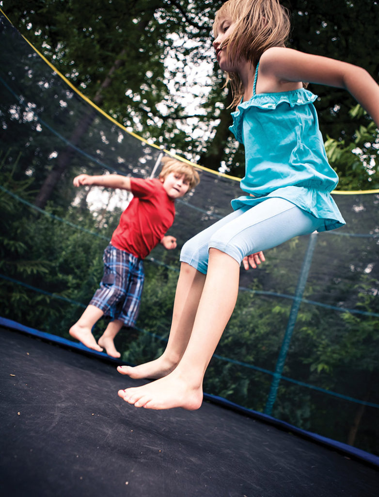
Image may be NSFW. Clik here to view.Trampoline use, both in backyards and in large recreational parks, is up. So are injuries incurred on the equipment, including fractures with potentially serious long-term sequelae. Some groups advise a ban on home use, but other experts disagree, citing the equipment’s benefits for motor learning and active play.
By P.K. Daniel
The late George Peter Nissen fashioned a canvas sheet to a rectangular steel frame in his parents’ garage nearly a century ago to create the world’s first bouncing apparatus, which he later called the trampoline. It’s doubtful, however, that he had in mind toddlers jumping or multiple children bouncing simultaneously, scenarios that often result in lower extremity injury.
Despite repeated warnings from the American Academy of Pediatrics (AAP) about the dangers of the trampoline, its popularity hasn’t waned. More than one million people suffered a trampoline injury resulting in an emergency department (ED) visit between 2002 and 2011, according to a national database study.1
“We see fractures from trampoline injuries almost every week in the summer months,” said Aaron J. Provance, MD, medical director of the Pediatric Sports Medicine Program in the Department of Orthopedics at Children’s Hospital Colorado in Aurora.
The national database study, conducted by researchers at Indiana University in Indianapolis, used the Consumer Product Safety Commission’s National Electronic Injury Surveillance System (NEISS) to look at trampoline-related fracture patterns across a large population. The results, published in 2014 in the Journal of Pediatric Orthopaedics, noted that 29% of the trampoline injuries were fractures, and more than a third (35.7%) occurred in the lower extremity.1
Tibia fractures often occur when a larger child is rebounding upward and a smaller child is landing simultaneously, causing significant compression force to the lower extremity.
Although 51.7% of those who suffered fractures were male, more female patients (54%) suffered lower extremity fractures. Nearly 93% of all fractures occurred in patients 16 years or younger (average age 9.5 years).1
There were nearly 105,000 ED visits in 2014 for trampoline injuries, according to Bob Segall, an investigative reporter at television station WTHR in Indianapolis, who in February reported on his analysis of NEISS data. In comparison, there were slightly more than 66,000 trampoline injuries requiring ED treatment in 1995.2
At least one study attributes the rise in injuries to an increase in trampoline sales, and, subsequently, in participation.3
Who’s at risk?
The type of trampoline injury varies with age and size, but the youngest, smallest kids are at the greatest risk.
“Younger and smaller children are much more likely to sustain fractures of all types,” said Michele LaBotz, MD, FAAP, a member of the national executive committee for the AAP’s Council on Sports Medicine & Fitness, who practices at InterMed in South Portland, ME.
The AAP reported that smaller, typically younger children are 14 times more likely to incur injuries than larger children.4
Segal at WTHR also reported the majority (85%) of the 2014 ED visits for trampoline injuries recorded in the NEISS database involved children; those aged 2 to 5 years accounted for a quarter of the visits. And 42% of the injuries suffered by toddlers and preschoolers were bone fractures.
Although the AAP and the American Academy of Orthopaedic Surgeons maintain that the majority of trampoline injuries occur in home environments, the proliferation of trampoline parks is another source of injuries. There was a staggering 700% growth in trampoline parks between 2011 and 2014, according to the International Association of Trampoline Parks, which itself was established only in 2012. These parks cater to kids—even those who are very young.
Many parks lack oversight. Arizona and Michigan are the only states with specific laws related to trampoline safety, and there are zero federal regulations for trampoline parks.
“With the increased number of companies having indoor trampolines, the injury rates may be similar to home-based trampolines,” said Provance. “Future research needs to be completed in this area.” LaBotz also said that, due to a lack of data, it’s unclear which setting–trampoline parks (with wall-to-wall and between-bed padding) or backyard home trampolines (some with safety nets)–results in more injuries.5
Anecdotally, however, LaBotz said sports medicine and ED doctors have noted marked increases in trampoline-specific injuries in geographic areas where trampoline parks have opened. “Visits to these trampoline parks often show an impressive list of rules and regulations upon entry, but enforcement of these policies [especially with regard to limiting numbers of jumpers on a mat] is highly variable,” she said.
Despite repeated warnings by the AAP that children younger than 6 years are at an increased risk of fractures and dislocations and should not use trampolines, trampoline parks offer programs, with such names as Rockin’ Tots and Toddler Time, specifically geared to this age group.
“[Toddlers’] lack of balance and muscle strength make them much more susceptible to more severe injury, especially in cases involving multiple simultaneous users,” said LaBotz. “Forces associated with rebound on the soft trampoline mat are far greater than forces that are generated by a fall onto hard ground, especially for smaller children.”
The multiple-jumper problem
About 75% of injuries occur when multiple people use the trampoline at once, according to a Consumer Product Safety Review, which the AAP reported in its 2012 policy statement and reaffirmed in 2015.4 When larger children are bouncing with smaller ones, they generate more recoil of the trampoline bed and more impact forces than smaller users can generate independently.4
Larger people on the trampoline bed sometimes “rocket propel” smaller children. In other words, they send the kids much higher in the air than they could go on their own. When they fall onto the mat, they fall from a greater height than they otherwise would, while the trampoline bed recoils upward at the same time.
NEISS database study coauthor Randall T. Loder, MD, chair of Indiana University’s Department of Orthopaedic Surgery, told LER: Pediatrics: “[In] my anecdotal experience taking care of many of these children, there is often more than one person on the trampoline, and there is the double-hit phenomenon—the child hits the trampoline when the other child is landing, doubling the force.”
Provance noted that tibia fractures commonly occur when a larger child or adult is rebounding upward and a smaller child is landing simultaneously on the trampoline, causing a significant compression force to the lower extremity. If this compression force is large enough, the top of the tibia can incur a buckle or compression fracture.
Injuries
Provance said most of the trampoline injuries he sees at the Sports Medicine Center at Children’s Hospital in Colorado are fractures of the upper and lower extremities. “The more serious injuries typically involve fractures at the top of the [tibia],” he said. “These fractures can have a risk of long-term complications. Even after these fractures have healed, there is a risk of further angulation of the leg months to years down the road.” 6,7
LaBotz said one of the main risks with these injuries is a lack of recognition. Presentation depends on the site of the injury. For example, physicians don’t typically look for a buckle fracture, which is an incomplete fracture in which one side of the bone is compressed, causing the other side to bend, in the proximal tibia, she said.
“Some of these injuries are rarely seen outside of trampoline-associated trauma, and providers might not be aware of the usual patterns of presentation,” LaBotz added. “One of the most common of these missed injuries is the metaphyseal fracture seen in the proximal tibia of young children. This is commonly a ‘torus-type’ of injury, which sometimes seems to escape recognition even by experienced radiologists, although once it is appropriately treated it heals well.”
Trampoline ankle
“Trampoline-related growth-plate fractures to the young ankle can be particularly severe and often require acute surgical intervention,” said LaBotz. “These injuries are at high risk for future growth arrest, which can be very problematic in the younger population.”
Paul Moroz, MD, a pediatric orthopedic and spine surgeon at Shriners Hospitals for Children in Honolulu, HI, agreed. He and colleagues addressed “trampoline ankle” in a study they conducted at the University of Ottawa in Canada and first published online in November 2015 in The Journal of Pediatric Orthopaedics.8
Moroz told LER: Pediatrics in its February issue that multiple jumpers are the primary cause of trampoline ankle (see “Multiple jumpers increase risk for trampoline ankle,” page 7). This injury involves the growth plates at the lowest part of the tibia and fibula, just above the ankle joint, making it unique to children.8
Just like the tibia fractures that Provance described as the result of compression forces, Moroz described how, when two individuals bounce out of sync, kinetic energy forces are generated that produce a high-impact effect. For youngsters this can cause serious distal as well as proximal growth-plate injuries that can result in long-term consequences, such as growth arrest. 6,7
Because there is a broad spectrum of injuries associated with trampoline use, treatments range from simple bracing or splinting performed in emergency or outpatient settings to injuries that require hospitalization and potential surgical intervention.
Image may be NSFW. Clik here to view.Improving safety
There doesn’t seem to be a difference in the severity or prevalence of injury with the use of padding, netting, or other safety measures. The AAP has reported that the introduction of netting and other safety equipment in the late 1990s and early 2000s has not reduced injury rates.4 Many injuries occur on the bed itself, where padding or netting can’t mitigate injury risk. In addition, LaBotz said many suburban neighborhoods have trampolines with nets and padding that are inappropriately installed, in poor repair, or both.
Another problem, she said, with “safety nets” is the often false perception of protection they can produce. They may, for example, entice users to take additional risks on the apparatus; falsely assure parents and other adults that supervision is not needed; and can be additional sources of injury, particularly if children try to climb netting or get body parts caught in the netting while jumping, all of which is why the AAP continues to advise against using backyard trampolines.
Not everybody is on the trampoline ban bandwagon, however. Norwegian researchers don’t support the AAP’s position. Instead, they advocate using the trampoline for lower extremity exercise to help children develop motor skills, strength, and balance.
In a 2006 issue of the British Journal of Sports Medicine, orthopedic surgeons from Trondheim, Norway, wrote: “Several reports on trampoline injuries recommend a ban on private, recreational trampoline use for children. We do not, for several reasons, support such a ban. Jumping on a trampoline gives children the ability to improve their motor control. It may also increase physical activity.”9
Although the Norwegian researchers stressed the importance of being aware of trampoline-associated risks, they concluded the effects of inactivity outweigh the risks of trampoline use.
LaBotz agreed with the Norwegians. She said there is an intrinsic risk associated with any form of physical activity—everything from bicycling to football can be associated with higher rates of acute injury compared with sitting on the sofa.
“But, if sitting on the sofa is all someone does, it will certainly contribute to one’s early demise over the longer term,” she said. “This risk-benefit balance will vary depending upon the nature of the activity itself, as well as of the population participating in it. Home trampolines are probably best compared to home swimming pools, in that they are very appealing and fun for many young kids, but, if not used appropriately and with adult supervision [supervision that is engaged and on site, not just peeking out the kitchen window], can be very hazardous. Our main concern at the AAP is not with trampolines per se, but with inappropriate use, and the current cultural perception and marketing of trampolines as toys.”
Athletes, including gymnasts, divers, figure skaters, freestyle skiers, and the like, use the trampoline as an intrinsic part of learning certain skills. However, this training is done with trampolines that differ from those used by the public; it also includes dedicated and experienced spotting and oversight that make the risks associated with this activity much lower than with recreational home trampoline use.
There are steps, however, that the US Consumer Product Safety Commission recommends to reduce serious injuries.4
They include: limiting use to one person at a time; not attempting flips; keeping springs covered with padding; not placing trampolines near trees or other structures; not allowing children younger than 6 years on the trampoline; providing adult supervision at all times; placing an enclosure to prevent falls to the ground; and not placing a ladder near the trampoline.
“Trampolines were initially designed for training acrobats and military aviators, and can be a very helpful part of a structured and well-supervised athletic training program, but they are not toys,” said LaBotz.
Although not directly related to the lower extremity, LaBotz emphasized that .5% of trampoline injuries result in permanent neurologic sequelae, such as paralysis or paresis to the upper or lower extremities or both resulting from cervical spine injury.10
“While this one-in-two-hundred rate may seem low, given the severity of consequences and the overall numbers of trampoline-related injuries, it is important to keep in mind,” she said. “The most common mechanism of cervical spine injury on the trampoline is due to failed attempts at aerial [front or back] somersaults or flips.”
The Indiana University study numbers confirm the frequency of trampoline injuries presenting to EDs, but the problem is more widespread, and the actual figures are likely much higher, LaBotz noted.
“This likely reflects significant under-reporting, in that many of these injuries [particularly with the high cost associated with ED care] will present to their primary care provider or to urgent care/walk-in other outpatient facilities for evaluation and treatment,” said LaBotz. “These injuries are not currently captured in NEISS or other large databases that are often used as information sources for these epidemiology studies.”
REFERENCES
Loder R, Schultz W, Sabatino M. Fractures from trampolines: results from a national database, 2002 to 2011. J Pediatr Orthop 2014;34(7):683-690.
National Electronic Injury Surveillance System (NEISS). 1995 NEISS data. Consumer Product Safety Commission website. http://www.cpsc.gov/en/Research–Statistics/NEISS-Injury-Data/. Accessed May 10, 2016.
Königshausen M, Gothner M, Kruppa C, Dudda M, et al. Trampoline-related injuries in children: an increasing problem. Sportverletz Sportschaden. 2014;28(2):69-74. [Epub 2014 Jun 25].
Briskin S, LaBotz M. Trampoline safety in childhood and adolescence. Council on Sports Medicine and Fitness, American Academy of Pediatrics. Pediatrics 2012;130(4):774-779. Reaffirmed July 2015.
Alexander K, Eager D, Scarrott C, Sushinsky G. Effectiveness of pads and enclosures as safety interventions on consumer trampolines. Inj Prev 2010;16(3):185-189.
Caine D, DiFiori J, Maffulli N. Physeal injuries in children’s and youth sports: reasons for concern? BR J Sports Med 2008;40:749-760.
Lalonde KA, Letts M. Traumatic growth arrest of the distal tibia: a clinical and radiographic review. Can J Surg 2005;48(2):143-147.
Blumetti F, Gauthier L, Moroz P. The ‘trampoline ankle’: severe medial malleolar physeal injuries in children and adolescents secondary to multioccupant use of trampolines. J Pediatr Orthop B 2016;25(2):133-137.
Nysted M, Drogset J. Trampoline injuries. Br J Sports Med 2006;40(12):984-987.
Brown PG, Lee M. Trampoline injuries of the cervical spine. Pediatr Neurosurg 2000;32(4):170-175.
Photo courtesy of Brian and Sarah Haigler, in memory of Maddy Haigler.
These children often have wider, more flexible, and more pronated feet than typically developing kids that don’t fit well into conventionally sized and shaped footwear. Ill-fitting shoes are linked to foot-specific disability and many other issues. Here, clinicians share strategies for finding the right fit.
By Lori Roniger
So many things aren’t designed for kids, who must make do in a world designed for relative giants. But most of them can enjoy getting a new pair of shoes in which they can walk, run and jump, and be raucous kids. However, finding footwear that fits well can be a challenge for the parents of children with Down syndrome.
In young children with Down syndrome, feet can be overlooked easily among the other challenges caregivers must handle, such as learning, cognitive, and dietary issues, and may not be a high priority, said Curt A. Bertram, CPO, FAAOP, national orthotics specialist and director of the National Residency Program at Hanger Clinic in Hartland, WI, who specializes in pediatrics.
The feet of children with Down syndrome often come with their own set of challenges. The dimensions of their typically wide feet tend not to conform to the conventional sizes in which most shoes are made. Their feet can be hypotonic, pronate significantly, and sometimes require ankle foot orthoses (AFOs) or other devices that can be difficult to fit in shoes. This makes it hard to find footwear in the proper sizes with appropriate support (see “Orthotic solutions for children with hypotonia,” LER: In Step with Pediatric Hypotonia, 2013, page 12).
Findings from an Australian study suggest children with Down syndrome may not be communicating foot-related problems to their parents effectively.
“You’re trying to fit a foot into a last or a shoe that’s made for a normal foot, and these feet are anything but normal,” Bertram said. “I tend to see them come in with shoes that are not the most appropriate.”
An article published in the Journal of Foot and Ankle Research (JFAR) last year that examined foot structure and footwear fit in 50 children and adolescents with Down syndrome found that ill-fitting footwear (often too narrow) was common and associated with foot-specific disability.1
Photo courtesy of Brian and Sarah Haigler, in memory of Maddy Haigler.
Additionally, flat feet were present in 76% of the study’s participants, hallux valgus in 10%, and lesser toe deformities in 12%. Hallux valgus was associated with foot-specific disability during school and play activities. The study also found foot structure and footwear fit were not significantly associated with parent-reported limitations regarding footwear choice, suggesting children may not be communicating foot-related problems to their parents effectively.
The study’s authors concluded that footwear education and regular footwear assessments could be helpful for children and adolescents with Down syndrome.
“In the daily grind of the whole host of other concerns of individuals with Down syndrome, footwear can be placed literally and figuratively down on the bottom,” said study author Nikolaos Nikolopoulos, BPod (Hons), MBusSys, LLM, a lecturer at La Trobe University in Melbourne, Australia, and a longtime clinical podiatrist.
In addition to ambulation and movement, footwear can also have implications for socialization and behavior, which can be important in children with Down syndrome, Nikolopoulos noted.
“Hopefully, we’re presenting a good case for footwear,” he said.
LER Pediatrics spoke with practitioners who work with this population about the challenges of finding footwear for children with Down syndrome that fits well and accommodates their unique needs.
Bertram recommended early intervention and assessment of the feet of children with Down syndrome. “Early” in this context means as soon as they start walking, which can occur later than in other kids, he said, and noted it’s better to treat them early to minimize the risk of foot pain and other foot problems as children mature.
“I think the tragedy that we want to avoid is [caregivers] thinking that they’ll outgrow it,” he said. “They won’t. They don’t. Their condition is permanent.”
Not just wider
Why can’t children with Down syndrome just wear wider shoes? Unfortunately, it’s not so simple.
“The Down syndrome foot tends to be short and wide and very, very, hyperflexible and pronated,” said David Jenkins, DPM, FACFAS, FAAPSM, professor at the Arizona School of Podiatric Medicine in Glendale. “Most shoes are not made for the pronated foot.”
Photo courtesy of Brian and Sarah Haigler, in memory of Maddy Haigler.
Collapsed arches can make the feet of children with Down syndrome wider, and shoes wide enough to accommodate their flat feet may not fit properly in other areas.
A lot of parents get shoes that are too long, which can cause difficulty when walking, said Faye McNerney, PT, DPT, C/NDT, who practices in Troy, OH. In the JFAR study, children’s shoes were, on average, 9 mm longer than their feet (range, 14.3 mm shorter-23.3 mm longer).
Orthoses, which require even more room in the shoes, can make footwear fitting more challenging. In the JFAR study, 38% of the children wore orthoses.
“A lot of children do have shoe-fitting issues, and we complicate this by giving them orthoses,” Bertram said.
The feet of children with Down syndrome also tend to have a medial bulge with a forefoot that abducts, he said. “Instead of a C-shaped footprint, the pronated foot looks like the opposite of that,” Bertram said.
Mismatched lasts
Jenkins has conducted research on the foot-to-shoe mismatch in more than 4000 Special Olympic participants with Down syndrome and other disabilities (Special Olympics does not track its athletes’ specific disabilities). Participants had a median age of 25.6 years, and some were as young as 5 years.2 Foot screenings found a significant 41% mismatch of feet to shoes, and a 20% referral rate of athletes for professional follow-up. Mismatch was defined as shoes that were 1.5 sizes too big or too small for the foot; 28.6% of athletes wore shoes that were too large, while 12.8% wore shoes that were too short.
“Conventional shoes available to all of us are not made on a last that’s appropriate for the Down syndrome foot,” Jenkins said.
Jenkins has also found that shoes that don’t fit contribute to secondary problems in this population, such as blisters, bunions, and ingrown toenails, as well as pain, disability, and increased body mass index.
“A lot of things had to be related to the shoe mismatch,” he said.
To that end, he is serving as a consultant to a company that is developing shoes designed for the feet of children with Down syndrome about 8 years and older. The company, Foot Options, hopes to introduce the footwear line during the fourth quarter of this year.
Shoe specifics
When a child with Down syndrome comes in for an assessment, Bertram first makes sure she or he has a good pair of shoes that fit properly. Next, if needed, is adding arch support to the shoes. After that, if the patient needs more control, he considers a supramalleolar orthosis (SMO) or an AFO.
Practitioners and researchers who focus on the feet of children with Down syndrome note that finding appropriate footwear for those who require a lot of support is in some ways easier than for the children who don’t wear orthotic devices. Children who wear AFOs or SMOs don’t require as much support from a shoe, though finding footwear that accommodates a wide Down syndrome foot, as well as a brace, can be challenging.
Photo courtesy of Brian and Sarah Haigler, in memory of Maddy Haigler.
An expensive shoe is not necessary, especially when kids are wearing an orthosis. “It’s basically a covering for the foot and orthosis,” McNerney said. A regular tennis shoe will do, and high-tops aren’t necessary.
“You still want a flexible forefoot, but you don’t need to have a particularly stable heel counter,” said Julia Looper, PT, PhD, associate professor of physical therapy at the University of Puget Sound in Tacoma, WA.
What is essential is for the footwear to fit over the device and stay put.
“Some parents buy cowboy boots,” McNerney said. “You don’t want that. You don’t want cute little church shoes.”
Even the thinnest braces can cause shoe-fitting problems and may require going up half a size, said Kathy Martin, PT, DHS, professor and assistant DPT program director at the Krannert School of Physical Therapy at the University of Indianapolis in Indiana and a pediatric physical therapist who works with children with Down syndrome.
For children who don’t wear braces, Bertram said a three-quarter top or high-top will provide better foot control than a low-top shoe.
“With hypotonia, you tend to have feet that don’t function the way they should,” Bertram said.
He recommends a stiffer shoe with solid construction, as well as a good heel counter and a wide sole, such as a high-quality athletic-type shoe.
“If you don’t need an orthotic, a high-top shoe can give support to the heel and ankle bone so the foot can’t roll in as much,” Looper said.
Martin added that a shoe with a straight or slightly curved last is better for these children, as a more curved last can encourage pronation.
“They should be [wearing] a more supportive shoe because they don’t have intrinsic ligamentous support in the foot,” she said.
Bertram advises that children with Down syndrome who have mild pronation and require arch support wear shoes with lacing rather than Velcro closures. Laces provide more support and control, though Velcro is preferable if the child wants to take the shoes on and off themselves.
McNerney, who is a big proponent of SMOs in children with Down syndrome, recommends shoes with flexibility in the toe box and emphasizes the importance of the right width.
Not only do shoes with laces provide a snugger fit than Velcro, McNerney said, she recommends them because, when kids are little, they can’t take them off when parents double-knot them. She noted this could otherwise be a problem when shoes are paired with a brace that takes some work to put on and secure properly in the shoe.
“I’ve heard this story a lot,” she said, regarding kids removing their shoes and braces while sitting in the back of the car.
But how do parents find shoes that are wide enough but not too long?
“The best advice is to keep going to different shoe stores,” McNerney said.
She said that some parents have gone to multiple shoe stores until they find the right shoes for their child with Down syndrome, and then they’ll buy the shoe in the next bigger sizes so they have it when they need it. She recommends that parents gravitate toward cheaper discount shoe and general merchandise stores.
“They typically carry the wide shoes, the rejects,” she said.
Looper suggested families learn which shoe brands work for their child’s feet, while Bertram cautioned that cost is not necessarily indicative of quality or shoes that control pronation.
“I think the parents definitely need to have direction, otherwise they do really have a hard time,” McNerney said about selecting shoes.
One trick Nikolopoulos recommended is tracing the foot on a piece of paper to provide a schematic of what is needed in the right shoe. Measuring both feet also can be helpful in this population, he said, particularly given that the children may be unlikely to report asymmetries in footwear fit that could become problematic.
Thinking long term
Although there isn’t yet any published research to prove it, some practitioners believe that not taking proper care of the feet of children with Down syndrome could cause secondary problems when they’re adults. Martin noted some adults with Down syndrome develop bunions—something seen in 10% of the children in the JFAR study—and overpronate.
Bertram said he sees blisters, calluses, and acquired deformities from shoe wear in teenagers and young adults who have Down syndrome. He said this can be due to wearing bad footwear that is too small or from uncontrolled deformities that were never managed. These anecdotal observations are consistent with studies indicating an association between hallux valgus and too-short shoes in typically developing children.
Bertram and Martin believe that individuals with Down syndrome are less likely to experience these secondary problems if, as children, they wear shoes that fit and provide support.
But, for now, children with Down syndrome still have imperfect options for finding well-fitting footwear that provides the support they need. And practitioners are doing their best to give their pediatric patients with Down syndrome and their caregivers sound advice about appropriate footwear.
Overall, though, practitioners like Martin concede they’re not the ones dealing with the kids every day, and that parents are better equipped to know if Velcro should win out over laces or if it’s reasonable to let their child wear cute sandals for a short amount of time.
“You pick your battles,” Martin said.
Lori Roniger is a freelance writer based in San Francisco, CA.
REFERENCES
Lim PQ, Shields N, Nikolopoulos N, et al. The association of foot structure and footwear fit with disability in children and adolescents with Down syndrome. J Foot Ankle Res 2015;8:4.
Jenkins DW, Cooper K, O’Connor R, Watanabe L. Foot-to-shoe mismatch and rates of referral in Special Olympics athletes. J Am Podiatr Med Assoc 2012;102(3):187-197.
Klein C, Groll-Knapp E, Kundi M, Kinz W. Increased hallux angle in children and its association with insufficient length of footwear: a community based cross sectional study. BMC Musculoskelet Disord 2009;10:159.
A recent study demonstrates the importance of personalizing prescriptions for ankle foot orthoses (AFOs) in children with cerebral palsy, rather than applying group data to individuals.
AFOs weren’t consistently associated with the study’s primary outcome, enhanced walking activity and intensity in a community-based setting compared with footwear alone. However, some individual children demonstrated significant improvements with AFOs.
“Orthotics are prescribed as individualized interventions,” said Kristie F. Bjornson, PT, PhD, associate professor of pediatrics and developmental medicine at Seattle Children’s Hospital in Washington. “While classical scientific inquiry uses group-based analysis, we know that, with children with cerebral palsy, none of them look exactly alike. We need to keep the individual level of analysis because that is what we do in clinical practice: prescribe on an individual level.”
Bjornson and colleagues conducted a randomized crossover study involving 11 children with CP (mean age 4.3 years, range 3-6 years) who typically wore bilateral AFOs. They randomized the children to either AFOs plus footwear for two weeks or footwear only for two weeks. Then each group crossed over to the other condition for another two weeks.
Individually, results of this real-world walking study varied, with no clear pattern for a child’s age or AFO type.
At the group level, the two conditions had similar results for average total strides per day (4660 strides, AFO-on; 4897 strides, AFO-off). Secondary outcomes, the percent of daytime hours walking, average number of strides faster than 30 strides/minute, and peak activity index, were similarly close.
Individually, however, the results varied, with no clear pattern for a child’s age or AFO type.
Two children (one aged 3.6 years with a solid AFO and a jump gait pattern, the other aged 4.3 years with a hinged AFO and a crouch gait pattern) took more steps per day with AFOs off than on. Another two children (one aged 3 years with a solid AFO and a true equinus gait pattern, and the other aged 3.6 years with a solid AFO and jump gait) took more steps with the AFOs on.
The two children whose steps per day improved with the AFOs on also spent more time walking with the AFOs on than off. Two more children (one aged 3.2 years with a solid AFO and a true equinus gait pattern, the other aged 5 years with a nonarticulated AFO and a true equinus gait pattern) demonstrated similar improvement. One of the children who improved in steps per day with the AFOs off (the older of the two) also spent more time walking with the AFOs off than on.
The same two children who improved in those two areas with AFOs on also had more strides per day at faster than 30 strides/minute and a higher peak activity index with AFOs on than off.
The pilot study results, published in the summer issue of Pediatric Physical Therapy, suggest most children weren’t wearing orthoses or footwear that helped them improve their daily walking activity in amount or intensity, Bjornson and colleagues wrote. They pointed out that the two children whose shank-to-vertical angle was optimized were also those who demonstrated the greatest positive effects of AFOs and footwear on daily walking activity and intensity.
“When they walked, they walked at higher rates,” Bjornson said. “Kids with CP walk slower and can’t keep up with their peers. So, hopefully, interventions would help them walk more, and, when they do walk, walk faster. Hopefully they can keep up with their peers walking to the bus, or they can run to first base when they want to when they play baseball.”
Elaine Owen, MSc, MCSP, a pediatric physical therapist at the Child Development Center in Bangor, North Wales, UK, told LER: Pediatrics the benefits for children with CP of walking outside with other children cover all areas of the International Classification of Functioning, Disability and Health, from body structure and functions to activities and participation.
“If they are walking with a good gait pattern they would be strengthening muscles, stretching muscles and joints, and [stimulating] motor learning, all things that will maintain or improve their physical condition,” Owen said. “Potentially, then, they may start to use less energy when walking and need fewer interventions. It may mean they have less pain in the teen and adult years.”
Bjornson is looking forward to more community-based research.
“Most orthotic research has been based on outcomes collected in a clinical or investigational gait lab. What a child does in front of somebody watching them is not necessarily what children do in their daily lives,” she said. “To my knowledge, nobody has ever looked at gait lab data compared to walking activity data. It’s a different kind of outcome.”
Larry Hand is a freelance writer in Massachusetts.
Source:
Bjornson K, Zhou C, Fatone S, et al. The effect of ankle-foot orthoses on community-based walking in cerebral palsy: a clinical pilot study. Pediatr Phys Ther 2016;28(2):179-186.
Overweight and obese children present with higher overall foot loading and a disproportional impact on the midfoot and longitudinal arch, with those as young as 1 or 2 years of age affected, according to research from Germany that may have implications for abnormal foot loading prevention in children.
Lead author Steffen Müller, PhD, research associate at the University Outpatient Clinic, Sports Medicine & Sports Orthopaedics, at the University of Potsdam in Germany, remarked, “In our data we cannot really see a well-adapted gait strategy for these children [for foot loading] since no real compensation is visible. Therefore, it could be speculated that the obese children are not capable [of adapting] their gait sufficiently to extra weight, leading to higher and disproportional foot loading.”
The study included 7575 children aged 1 to 12 years and examined foot-loading characteristics during gait. Of the participants, 6458 were categorized as normal weight, 746 as overweight, and 371 as obese, according to a German reference system based on age- and gender-specific body mass indices.
Plantar pressure measurements were taken during gait with a pressure measurement platform that was mounted on an instrumented walkway. The researchers calculated the contact area, arch index, peak pressure, and force time integral for the total foot, forefoot, midfoot, and hindfoot.
Weight-related differences in peak plantar pressure, while relatively small, were present at the mid- and forefoot by age 2 years.
The data suggested a mean walking velocity of .95 m/s was evident in normal-weight, overweight, and obese children. However, obese and normal-weight children presented with the highest and lowest foot contact area, respectively, in all age groups. This was also the case for the arch index in children aged 5 to 12 years.
Müller suggested that prevention strategies, such as strengthening and sensorimotor exercises for the foot and lower extremity, should start at a young age. “Additionally, we should try to enhance the compliance of these children to participate in an active lifestyle and reduce weight,” he added.
Meanwhile, overall foot loading and peak pressures increased consistently with age and were highest in the obese children and lowest in the normal-weight children. Increased foot loading for the total foot and forefoot was present from the age of 3 to 4 years, while excess foot loading at the rearfoot and midfoot was visible from age 5 to 6 years.
Müller said, “Since pros and cons for active versus passive treatments in the long run are well known, active interventions should be preferred in contrast to shoe and insole adaptations to address disproportional foot loading and/or foot stress/pain.”
The differences in peak pressure at the toes, rearfoot, and total foot were relatively small between normal-weight, overweight, and obese children, but were present at the forefoot and midfoot at age 2 years. PLoS One published the findings in February.
Commenting on the study data, Sharon Bout-Tabaku, MD, assistant professor of pediatrics at Nationwide Children’s Hospital in Columbus, OH, said: “The implications are important because we can hypothesize that walking speed, lower extremity strength, and altered forces around the knee and hips will be affected.”
She added that, “Early foot changes may affect the morphology of the bones and joints due to sustained low-level systemic and local joint inflammation. Finally, we can guess that it may interfere with optimal physical function and can be associated with pain.”
She also emphasized weight management. “Certainly maintaining a healthy weight or weight loss early is important because it can minimize alterations in morphology, forces, and gait across the entire lower extremity,” she said. “Physical activity and normal childhood play is equally important to maintain bone development, strength and neuromuscular development.”
She added that “in children who are symptomatic, orthotics, heel cups, and cushioning may provide some relief.”
The study authors concluded the plantar pressure values assessed in this trial could serve as reference data to identify suspicious foot-loading patterns in children. Bout-Tabaku agreed, and noted, “This article provides nice reference data in a large sample and serves as a starting point for other studies: longitudinal studies; studies exploring associations with foot pressure and function, gross motor skills, and pain; and longitudinal studies looking at bone and joint development radiographically.”
Bout-Tabaku added that “the take-home is that obesity is modifiable. Similarly, during childhood and adolescence, the musculoskeletal system is dynamic and very responsive to change, and can be positively modified with weight loss in childhood. The degree of positive changes in the musculoskeletal system is harder to attain with increasing age.”
Katie Bell is a freelance writer based in New York City.
Sources:
Müller S, Carlsohn A, Müller J, et al. Influence of obesity on foot loading characteristics in gait for children aged1 to 12 years. PLoS One 2016;11(2):e0149924.
Image may be NSFW. Clik here to view.Safety of underhand pitching overrated
By Hank Black
Better pre- and offseason lower-body conditioning may help prevent some serious overuse injuries to the upper extremities of young female fast-pitch softball players, according to the lead author of a recent prospective study of player-reported injuries.
“Because the lower body is the driving force of the windmill pitch, a good conditioning program for the gluteal musculature would be important in sequentially stabilizing the pelvis and trunk to provide support for the rest of the kinetic chain,” said Matthew V. Smith, MD, lead author and assistant professor in the Department of Orthopedics at Washington University in St. Louis, MO.
The study included 98 select-level players: 48 pitchers and 50 position players aged 9 to 18 years. Investigators used a web-based survey to gather information on self-reported injuries during a single April to August season. They defined injury as pain that sidelined a player for the remainder of the current practice or game or from her usual participation the day after injury.
Forty-three of the players reported 49 injuries, with a cumulative incidence of injury of 40% (this includes 58 athletes who didn’t respond to the survey); 70% of injuries were to the lower extremities.
The injuries that resulted in loss of significant playing time, however, were primarily to upper extremities and incurred by pitchers. Pitchers sustained 30 of the injuries; 18 of these were directly attributed to pitching.
“Positional players had the standard lower extremity sprains and contusions you’d expect to see in most sports and that kept them from participation for less than two weeks; these were, for the most part, unpreventable,” said Smith. “In pitchers, however, severe shoulder injuries, as well as some stress fractures in the forearm, prevented participation for more than two weeks and appeared to result from overuse in an environment where they pitch in two or three games a day in tournaments lasting up to four days.”
The majority (78%) of pitching injuries happened in the first six weeks of the season, so an increased emphasis on offseason and preseason conditioning may be advisable, said Smith. He also noted the number and severity of pitching injuries counter the notion that fast-pitch softball’s underhand windmill pitch is safe relative to baseball’s overhand pitch. “Underhand pitching is not as safe as some think it is,” he said.
In an ongoing follow-up study, the researchers are using motion-capture technology to gather data on pitchers’ movements during simulated games, as well as collecting ground-reaction force data.
“We are evaluating what happens to lower extremity mechanics as pitchers tire. Our hypothesis is that, as throwing athletes fatigue in their arm and shoulder, their lower body mechanics will change in an attempt to maintain velocity. For example, trunk rotation or pelvic tilt may change to try to use more components of the kinetic chain to generate arm speed,” Smith said.
“Many athletes of this age may get hitting or pitching instruction, but not a good conditioning program to get them ready for the intensity of their sport when the season starts. I think that’s a really critical thing parents should push for–not so much performance training but performance conditioning,” he said.
Sports Health published the study in its November-December 2015 issue.
“The paper is correct in citing studies that show softball pitching is no easier on the body than baseball pitching,” said Gretchen D. Oliver, PhD, FACSM, ATC, director of the Sports Medicine and Movement Laboratory at Auburn University in Alabama. “Both baseball and softball pitchers will incur injury if their mechanics are flawed or inefficient.”
Oliver, who is an associate professor in the School of Kinesiology, agreed that lower body mechanics of softball pitchers are important to prevent injury.
“Proper utilization of the kinetic chain allows for efficient kinetic energy transfer from the proximal segments to the distal segments. Dysfunction at a proximal segment may lead to altered energy transfer and dysfunction at more distal segments,” she said. “We emphasize total body conditioning and focus a lot on postural control activities. The more body awareness a pitcher has, the better chance they have to develop consistency in their mechanics.”
She noted, “The study did not examine preseason conditioning, so that could be a factor in the early-season injuries, but there are many other factors that should be examined before we can determine the ‘why.’ We are now tracking pitch volume yearlong, as well as examining performance measures.”
Hank Black is a freelance writer in Birmingham, AL.
Source:
Smith MV, Davis R, Brophy RH, et al. Prospective player reported injuries in female youth fast-pitch softball players. Sports Health 2015;7(6):497-503.
 Some report more pain,