![Photo by Joshua Albanese (joshuaalbanese.com)]()
Photo by Joshua Albanese (joshuaalbanese.com)
Early diagnosis and early intervention in people with Charcot-Marie-Tooth disease give clinicians and patients a valuable head start on keeping symptom progression in check.
By Cary Groner
David Misener, CPO, understands Charcot-Marie-Tooth (CMT) disease firsthand because he has it himself, as does his son. Misener, who practices with Clinical Prosthetics and Orthotics in Albany, NY, said his case was difficult to diagnose at first because he and those in his family don’t carry the genetic mutation usually associated with the disease.
“They first ran the test on CMT1A, because that’s the most common, but they didn’t find the error,” he told LER.
As it happens, his family carries a different genetic mutation, CMT1B, which has the same symptoms but originates in a different gene.
“If you’re not one of those with type one-A, they have to keep looking, and it can become expensive,” Misener said.
Closing the information gap
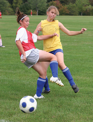
CMT, a sensorimotor neuropathy marked by progressive muscular atrophy, was first described in 1886 by physicians from France and England. The disease typically begins in the intrinsic muscles of the foot, then extends to the peroneus brevis and longus, the tibialis anterior, the extensor digitorum longus, and the extensor hallucis longus. It may eventually involve the hands, as well, and it’s even been associated with seemingly unrelated conditions such as scoliosis.1,2 Roughly one in 2500 people in the US has CMT, according to the National Institute of Neurological Diseases and Stroke (NINDS), and men are affected more often than women, in approximately a 5:3 ratio.3
Because CMT affects both motor and sensory nerves, patients can experience not only muscle weakening and imbalance, but also problems with proprioception.
David Misener’s diagnostic challenges occurred 20 years ago, and since then researchers have continued to refine their understanding of the genetic mutations that cause CMT. Research has revealed ever-expanding subclasses of the disease, in fact, which is usually inherited in an autosomal dominant pattern (ie, if one parent has CMT, each child has a 50/50 chance of inheriting it). However, some CMT subtypes may be passed on in an X-linked, or recessive, pattern, and the disease may also arise due to spontaneous or de novo mutations.4
“The most common subtype, CMT1A, affects about half of CMT patients,” said Michael Shy, MD, a professor of neurology, pediatrics, and physiology at the University of Iowa’s Carver College of Medicine in Iowa City. “As of now, four subtypes account for ninety percent of genetically identifiable CMT: CMT1A, CMT1B, CMTX, and CMT2A.”
According to Shy, CMT1A is considered the classic phenotype.
“These patients are likely to have been slow runners and clumsy as children, and as they reach adulthood many will need foot orthoses or ankle-foot orthoses (AFOs) to help them ambulate,” he continued. “Only a small percentage will ever need a walker or a wheelchair.”
Other subtypes exhibit different characteristics, Shy said. For example, some CMT1B patients don’t begin to walk until two or three years of age and are severely affected in the first two decades of life, as is also the case with many CMT2A patients.
“It just depends on the particular mutation within the subtype,” he said. “It can range from severe early on to very mild, though as a rule the recessive types—such as CMT4—are often fairly severe.”
![Photo by Jordana Bieze Foster.]()
Photo by Jordana Bieze Foster.
Pathophysiology
CMT pathophysiology has traditionally been classified as either predominantly demyelinating or axonal, but clinicians and researchers disagree about the relative importance of these categories with regard to manifestation and progression of the disease.2 For example, although axonal degeneration is a predictor of disability—suggesting that axonal damage may be the root cause of the neuropathy—the mutations responsible for the different forms of CMT1 typically occur in myelin genes, and myelin disturbances lead to axonal damage. Further muddying the waters is that axonal damage can result in secondary demyelination.2
Genes cause CMT by disrupting particular proteins, Shy explained, and if those proteins form a constituent of myelin, the myelin will be damaged.
“You will see axonal damage in CMT1,” he said, “but the primary problem is the myelin.”
Some aspects of CMT remain perplexing, however.
“Many of the genes that cause CMT2—the axonal form of the disease—are expressed widely throughout the body, not just in neurons,” Shy said. “So why other organ systems aren’t affected is a mystery. It probably has something to do with the biology of the neurons, since they don’t divide, and have long axons that have to be maintained throughout the person’s lifetime.”
Axon length likely explains why distal parts of the body, starting with the feet, are affected first. And, because CMT affects both motor and sensory nerves, patients can experience not only muscle weakening and imbalance, but also problems with proprioception, Shy said.
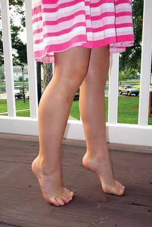
Some muscles weaken more quickly than others, moreover, which leads to imbalances between dorsiflexors and plantar flexors. These imbalances, in turn, contribute to contracture of the Achilles tendon and problems such as cavus feet, calcaneal inversion, forefoot adduction, and claw toes.5 Other CMT-related problems include skinny calves, scoliosis, foot drop, “slapping” gait, foot numbness, muscle weakness, balance difficulties, and multiple non-neuropathic pain symptoms.2,4
Diagnostic challenges
The heterogeneity of symptoms associated with CMT, and the subtlety with which they often present, may lead to delayed diagnosis in some patients.
“Many times people get diagnosed well after the age of onset,” David Misener said. “Those with mild cases may not realize they have the disease until they’re in their sixties and develop a drop foot.”
Because more than 80 CMT subtypes have been identified, the broad range of severity and age of onset can also contribute to missed diagnoses.
![Photo by Vincent Giordano Trinacria Photography (trinacriaphotography.com)]()
Photo by Vincent Giordano
Trinacria Photography (trinacriaphotography.com)
“In many families with CMT, you sometimes reach a tipping point where something gets your attention, such as stumbling,” Misener continued. “I think the disease probably progresses at an even rate, but certain events make you think it may have advanced more suddenly.”
Michael Shy agreed.
“In CMT1A patients, the disease tends to develop slowly through the first two decades of life,” he said. “Some CMT1B can present early, others don’t present until adulthood. It depends on the particular mutation.”
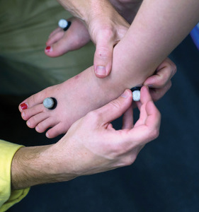
In making a diagnosis, Shy typically looks for length-dependent weakness and sensory loss, particularly in the hands and feet.
“Usually we look for absent ankle reflexes,” he said. “In some cases, all the deep tendon reflexes can be absent, and then we use nerve conduction testing to determine whether or not it’s primarily a problem with myelin or the axons.”
Although Shy and other specialists are attuned to CMT, many clinicians are not. The result, he said, is that the initial diagnosis is sometimes made by a foot specialist rather than a primary care physician.
“I think podiatrists are often the first people to recognize that someone may have CMT, because they see pes cavus or other foot abnormalities,” Shy said. “Length-dependent weakness and atrophy, and decreased deep-tendon reflexes are things that primary care physicians may pick up, though.”
Ken Cornell, CO, who practices with Cornell Orthotics & Prosthetics in the Boston area, finds himself making the initial diagnosis less often than he used to.
Patients usually present with one or more of five key symptoms: poor balance, foot drop, lateral ankle instability, sensory loss, or painful calluses.
“Twenty or twenty-five years ago I was getting a lot of these patients, and I’d call their doctors to discuss CMT, and they wouldn’t even know what I was talking about,” he said. “In the last few years that’s started to change, I think partly due to outreach from the CMT Association.”
Cornell said his patients usually present with one or more of five key symptoms: poor balance, foot drop, lateral ankle instability, sensory loss, or painful calluses. And, although nerve conduction studies have typically been considered definitive, increasing knowledge about the genetic basis of CMT has elevated demand for genetic testing.
![Photo by Vincent Giordano Trinacria Photography (trinacriaphotography.com)]()
Photo by Vincent Giordano
Trinacria Photography (trinacriaphotography.com)
Disease progression
Once the diagnosis of CMT is made, through either traditional means or genetic testing, clinicians and patients must monitor the disease’s progression to determine the most appropriate interventions. As noted previously, the disparity in weakness between opposing muscles may ultimately lead not just to imbalances but to deformities.
“The longest nerves in the body are affected first,” reiterated Ken Cornell. “In a man who’s six feet tall, one single nerve cell going from the spine to the foot can be three feet long. That cell is affected first, and that in turn has effects on the muscles that extend the toes. But the muscles that flex the foot are higher up in the leg; they’re shorter nerves, affected later, so you wind up with unopposed toe flexion. That’s how you get claw toe deformity.”
“The nerves are not feeding the muscles, so they start wasting away, and this creates muscle imbalances over specific joints,” added David Misener. “As one muscle pulls harder over that joint, it can affect the shape of the foot, which becomes cavus. Drop foot arises because the anterior muscle group weakens before you reach that tipping point in the calf muscle, and those anterior muscles lift the foot during swing phase and decelerate it at heel strike. These things start with the small intrinsic muscles in the feet, then affect the ankles, then the knees, and in some people, the hips.”
Cornell added that these disease traits make progression reasonably straightforward to predict.
“People are typically focused on the motor loss first, which usually manifests first with foot drop, then with everter paresis,” he explained. “The peroneus longus—the plantar flexor of the first ray—outlasts its antagonist, the anterior tibialis, so you wind up with a plantar flexed first ray and a tripod effect. This causes an inversion twist to the rearfoot, which sets you up for lateral ankle instability and forefoot adduction. All these muscle imbalances are progressive, and they set up the conditions for deformities.”
Michael Shy noted that certain types of nerves seem to develop problems more quickly than others, and it may not always have to do with nerve length.
“For reasons that are unclear, the nerves that cause dorsiflexion and eversion seem to get damaged earlier than those that cause plantar flexion and inversion, so you get a lot of anterior calf wasting,” he said.
With all the attention to motor nerve deficits, Shy added, CMT’s effect on sensory nerves shouldn’t be downplayed.
“Most CMT types affect both motor nerves and the large-fiber sensory peripheral nerves, which tell our brain where we are in space,” he said. “This can cause problems with balance. Some forms of CMT affect only motor nerves, others affect primarily sensory nerves, and why that difference exists isn’t completely understood yet.”
Interventions
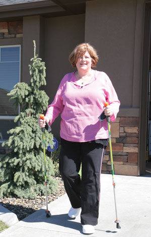
Although CMT isn’t curable, interventions such as physical therapy, exercise, and orthotic device use can ameliorate its effects.
“We recommend stretching and moderate-intensity exercise for CMT patients,” said Katy Eichinger, DPT, CS, who practices in the neuromuscular division of the Department of Neurology at the University of Rochester in New York. “Patients should be evaluated by a physical therapist, who can guide them to a program that’s best for them. Assessing a person’s strengths, weaknesses, age, disease progression, and personal desires can make an individualized program much more successful.”
![Photo by Joshua Albanese (joshuaalbanese.com)]()
Photo by Joshua Albanese (joshuaalbanese.com)
In terms of stretching, Eichinger emphasizes the ankle plantar flexors (a calf stretch, facing the wall with the leg extended backward and the heel on the ground, is a typical approach). For strengthening, she works with patients’ likes and dislikes to find something they can stick with.
“You can optimize patients’ functional ability and make sure no weakness occurs due to disuse or aging,” she explained. “If someone doesn’t like gyms, they may be willing to do aquatic exercises or ride an exercise bike at home.”
Because CMT patients have distal weakness, physical therapy should focus exercise programs more proximally, according to Eichinger. This means an emphasis on the quadriceps, the hip muscles, and the core. It also may mean balance training, particular in those whose neuropathy forces them to rely more on their vision and vestibular systems to maintain an even keel.
Where we go from here
With improved genetic sequencing techniques and increased understanding of proteins and myelination, researchers are moving closer to finding a cure for CMT. Today, the best treatments for CMT include regular exercise, maintaining a healthy body weight, and, when necessary, the use of orthotic devices. Approaches to orthotic management are discussed in detail in the article that follows.
Cary Groner is a freelance writer in the San Francisco Bay Area.
REFERENCES
- Groner C. Orthotic management of Charcot-Marie-Tooth. LER 2013;5(5):29-38.
- Gondim F. Hereditary neuropathies of the Charcot-Marie-Tooth disease type. Medscape website. http://emedicine.medscape.com/article/1173484-overview. Updated March 20, 2014. Accessed March 24, 2015.
- NINDS Charcot-Marie-Tooth disease information page. National Institute of Neurological Disorders and Stroke website. http://www.ninds.nih.gov/disorders/charcot_marie_tooth/charcot_marie_tooth.htm. Accessed March 24, 2015.
- Charcot-Marie-Tooth Association website. http://cmtausa.org. Accessed March 24, 2015.
- Kamp JJ. Orthotic management of Charcot-Marie-Tooth. J Prosthet Orthot 1994;6(4):108-112.

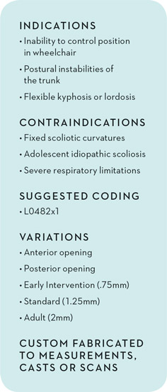 SureStep TLSO
 SureStep Criss Crossers
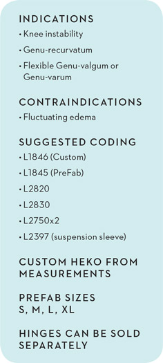 SureStep HEKO Custom
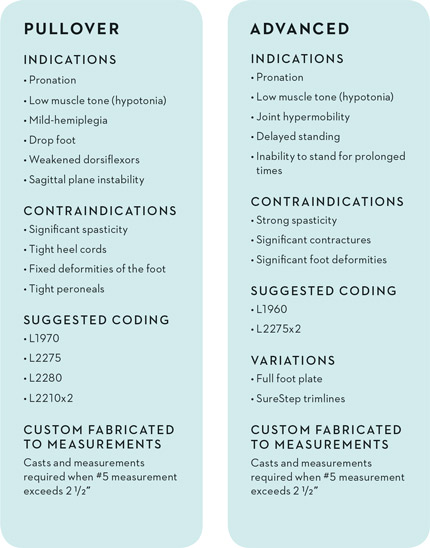 SureStep PullOver
 SureStep Indy 2 Stage

 SureStep BigShot

 SureStep SMO


 Location of shortening is key factor
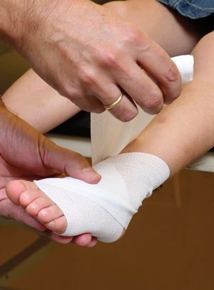 Experts push for better sprain rehab
 While the patient is always the practitionerʼs first priority, establishing a convivial relationship with a childʼs parents can mean the difference between victory and failure when it comes to diagnosing, treating, and rehabilitating lower extremity problems in pediatric patients.
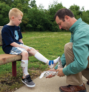


 By Shalmali Pal

 By P.K. Daniel
 Taking a conservative approach to returning to play seems to be gaining popularity among adults, as well. Minnesota Timberwolves point guard Ricky Rubio went down with a severely sprained left ankle on November 7, 2014. Rubio remained sidelined until February 2, frustrating T-Wolves fans. But Rubio’s recovery process was delayed—perceived instability in his ankle or not—because of muscle and ligament damage. Minnesota coach Flip Saunders said two medical specialists who consulted with Rubio advised that returning too soon could increase the risk of a stress fracture.
 Sole stiffness affects stance time
 Experts call for age-specific options


 Once pain and inflammation have been addressed, clinicians can implement interventions—including orthotic devices, stretching, and strengthening—to address the biomechanical factors that are believed to contribute to heel pain and other symptoms in this population.