Quantcast
Channel: Special Section – Lower Extremity Review Magazine
Viewing all 184 articles
Browse latest View live

Lean manufacturing transforms orthotic fabrication

$
0
0

CONFERENCE COVERAGE: Orthotics Technology Forum 2013

By Emily Delzell

The need to replace entrenched processes and thinking with fluid, fast-moving orthotic design and manufacture that minimizes errors and maximizes resources was highlighted by several speakers at the Orthotics Technology Forum, including Jarret Eschenburg, CPed, director of operations at Coral Springs, FL-based SureFit, a subsidiary of Hanger.

Eschenburg made a distinction between simply “having technology” versus “using technology” in an optimal way—one that facilitates continual change and improvements in orthotic fabrication and results in an enhanced bottom line for lab owners.

From clutter to clean: The tracing library before and after SureFit’s process-flow kaizen. (Photos courtesy of Jarret Eschenburg, CPed.)

From clutter to clean: The tracing library before and after SureFit’s process-flow kaizen. (Photos courtesy of Jarret Eschenburg, CPed.)

This effort to make the most of expensive technology—SureFit invested in CAD-CAM in 2005, adding two mills to its lab—resulted in a reengineering process that streamlined the fabrication workflow and reduced workforce from 30 to 17 and turn-around times from 14 days to seven.

With new technology in place, Eschenburg continued his quest for kaizen, a Japanese term for continuous improvement, by embracing lean manufacturing, defined as “a manufacturing philosophy that shortens the time line between the customer order and the product shipment by eliminating waste using continuous improvement techniques.”

The first step to getting “lean,” he said, is standardizing work. A review of SureFit’s lab processes showed each employee did the same task slightly differently, from the orthotic gluers and grinders to the certified pedorthists who analyzed orders. The review also identified sources of waste, Eschenburg said, which in orthotic fabrication often involve quality issues, such as reworking orders, and workstation delays that cause bottlenecks.

“Wait times between processes eat up time. You may say it takes only about 45 minutes to actually make an orthotic, when in reality, it takes all day,” he said.

“Key lean concepts are using your current resources, and changing from the bottom up, not from the top down. It’s hands-on workers who really know what the issues are and can best suggest ways to improve,” he said. “Once processes are standardized you can implement crosstraining in which, ideally, every worker can do every job.”

Eschenburg next focused his continuous change efforts on three areas in orthotic manufacturing: reducing distances traveled by reducing the lab’s footprint, standardizing jobs to increase speed, and setting up a pull system to lower WIP (work in process) in the target area (postmill to grind finish).

“When orders get backed up you have increased WIP and work time. At this time we had a WIP of 400 orders—when I said we were aiming for 20 we almost had a revolution. We had a two-day turnaround in that area; we wanted it done that same day,” he said.

Every employee took part in the brainstorming that resulted in a new floor plan and a standard workflow for every operator—about 175 action items in all.

It took about three months to identify and implement changes, but the end results were worth it, Eschenburg said. SureFit reduced its lab footprint by 30% and achieved other efficiencies, such as setting up a standardized gluing process and simplifying tracing (see figure above).

“I regard SureFit’s progress merely as a means to do more. It’s an indication that we have reached a place where we might begin to improve again,” Eschenburg said.


3D printing: The shape of things to come

$
0
0

CONFERENCE COVERAGE: Orthotics Technology Forum 2013

By Emily Delzell

The Orthotics Technology Forum presentation given by Ben Boyer, CPed, was as much about what the speaker was wearing as what he was saying. Boyer, who is the lab manager at Kintec in Vancouver, Canada, wore a product he thinks may represent part of the future of orthotic design and manufacture—orthoses he’d printed with a hobbyist-level desktop 3D printer.

Boyer assured the audience that 3D printing (also called rapid prototyping, additive manufacturing, and mass customization) has become more accessible and cost effective, and will soon change the orthotic industry in the same way digital technology has changed media consumption.

“3D printing takes a physical object and makes it a digital commodity that anyone can produce anywhere, anytime,” said Boyer, who made his orthoses using a free downloadable project from thingverse.com. The site allows users to share CAD designs, which others can download, customize, and print.

Boyer was quick to note his printed devices, designed by a layperson, were “pretty horrible” and coming apart after a few months.

“The life span of materials used in 3D printing can be short, and materials that work well for production scale are currently cost-prohibitive,” he said, but added, “It is a product and may be good enough for some patients to try. And it’s only going to get better.”

Large labs will use 3D printing to improve production efficiency, he predicted.

“There will be no grinding, no cutting, it’ll come out and you’ll add a topcover, and that’s it,” he said. “As multimaterial printers get more sophisticated you’ll be able to print the shell, padding materials, and topcover all a once.”

In his presentation Boyer discussed podiatrist James Woodburn, MPhil, PhD, FCPodMed, professor of rehabilitation sciences at Glasgow Caledonian University in Scotland, who is now using a 3D printer in his clinic to print orthoses and other devices.  Woodburn is also leading the A-FOOTPRINT project, an international consortium that will commercialize fully integrated, cost-effective 3D-printed foot orthoses with a 48-hour manufacture time.

Chris Lawrie, Delcam’s business development manager for healthcare, pointed out that Woodburn had attended the first OTF, held in 2011 in Bath, UK, and expressed interest in an early model 3D printer.

“This shows the technology forum works,” said Lawrie, who noted it’s the exchange of ideas in settings like the OTF that puts advances into practice.

Woodburn’s current printer cost about $3000, which Boyer says is typical for a consumer-level machine. Production-grade printers can cost anywhere from $10,000 to $600,000.

Where does all this leave labs and practitioners who make orthoses?

“Maybe it’s a change in what we do,” Boyer said. “We will still do the clinical work, but then perhaps provide a high-quality design file and sell that. We can also provide follow-up care when patients do make their own orthoses.”

Other paradigms might include creating apps that make orthoses, he said.

High-tech path leads to the future of improved orthotic prescription

$
0
0

CONFERENCE COVERAGE: Orthotics Technology Forum 2013

An image of the future shown at the 2013 OTF: A 3D-printed model of the human foot, part of the A–FOOTPRINT project (see “3D printing,” page 40). (Photo courtesy of Peter Devlin.)

An image of the future shown at the 2013 OTF: A 3D-printed model of the human foot, part of the A–FOOTPRINT project (see “3D printing,” page 40). (Photo courtesy of Peter Devlin.)

By Emily Delzell

One of the most-talked-about images from the 2013 Orthotics Technology Forum (OTF) depicted 11 custom foot orthoses made by 11 experienced orthotists and podiatrists for a single patient. All 11 practitioners had been given the same information about the patient’s condition, yet all 11 orthoses were distinctly different (see figure, page 40).

The image, part of a presentation by Nachiappan Chockalingam, MSc, PhD, CEng, CSci, underscored the opportunity for advanced technology to take the guesswork out of orthotic prescription—a theme that was repeated throughout the OTF, which was held in late May at the Georgia Institute of Technology in Atlanta.

More than 80 attendees enjoyed several days of presentations, conversation, and a tour of Georgia Tech’s clinical biomechanics labs and the associated Global Center for Medical Innovation. In addition to host university Georgia Tech, event sponsors included Delcam Healthcare Solutions, Freedom Machine Tool, nora systems, SureFit, Mile High Orthotics Lab, Walking Mobility Clinics, Acor, Kiwi, and Kintec.

Drilling down into foot function

Chockalingam, professor of clinical biomechanics at Staffordshire University in Stoke-on-Trent in the UK, and others in his lab are combining plantar pressure and imaging data to develop patient-specific modeling to make orthotic prescription more accurate.

“[We can] provide more detail about foot function through mathematical models that could keep things like this from happening,” he said, pointing to the image of the 11 different orthoses.

Differences in orthotic training and approach result in highly variable prescriptions using raditional methods. (Image courtesy of Nachiappan Chockalingam, MSc, PhD, CEng, CSci.)

Differences in orthotic training and approach result in highly variable prescriptions using raditional methods. (Image courtesy of Nachiappan Chockalingam, MSc, PhD, CEng, CSci.)

His team looks at both static and dynamic measurements using motion-capture systems and marker sets; force plates; in-shoe and mat plantar pressure systems; and systems that use interdigital sensors to measure dynamic plantar pressure. They also use ultrasound and magnetic resonance imaging to look at changes in foot tissue under different conditions.

“We also use multisegmental foot models to measure relationships between different foot segments rather than measuring simple angles and range of motion,” he said. “For example, we’ll look at the relationship between the medial forefoot and the lateral forefoot, the forefoot and the rearfoot, the rearfoot and the shank.”

Chockalingam and his colleagues have developed a device that combines ultrasound with dynamometry to measure mechanical response to plantar loading. His team developed this as part of its involvement with DiaBSmart (development of a new generation of diabetic foot­wear using an integrated approach and smart materials), an international effort funded by the EU.

They found normal pressure differential gradients were 6.25 times higher in patients with diabetes than in controls, a factor that may be an independent predictor in diabetic foot ulceration, Chockalingam said.

“Physiological changes can affect mechanics, and various technologies can help us develop patient-specific and predictive models that can alter the orthotic prescription and process,” he said.

A closer look at tissue behavior, stiffness

In his Georgia Tech lab, research scientist Géza F. Kogler, PhD, CO, is also combining imaging and pressure measurement tools to produce unique data on tissue behavior and stiffness characteristics of the foot.

“There are all these different soft tissues through which you’re having to transfer a load [when an orthosis is used], and if I want to transfer a load I need to know what those tissues are and what their different compositions and mechanical properties are,” he said in an OTF presentation.

Kogler, who is director of Georgia Tech’s Clinical Biomechanics Laboratory, and his team have developed test apparatuses to quantify plantar midfoot soft tissue stiffness properties and behavior in nonweight-bearing and weight-bearing conditions to provide perspective on the diversity of tissues.

“We had a fairly simple hypothesis; that plantar midfoot tissues would be stiffer in weight bearing than in nonweight bearing because, when we load the foot, its structures become tense,” he said. “Most tissue studies have been done at the heel, but most of the control in an arch mechanism is at the midfoot, so we started there.”

They took measurements from a halfway point between the metatarsal heads and the posterior aspect of the calcaneus under weight-bearing and nonweight-bearing conditions, using ultrasound to quantify tissue displacement.

“We proved our intuitive hypothesis, that tissues were stiffer under weight-bearing than nonweight-bearing conditions, and also saw that tissue behavior and stiffness varies in individuals due to BMI and individual differences in tissue structure,” said Kogler, who had tested 25 healthy volunteers at the time of the OTF (the study is ongoing).

“What we’re really after is the clinical application [of this research], and to get to where we can determine what that stiffness value is in a particular foot and how we might use that to our advantage to control the movement with an orthosis, which means we need to alter the shape and perhaps the density of the material,” he said.

Kogler also discussed pilot study data that showed an orthosis changed the stiffness properties of plantar tissues compared with a barefoot condition.

“We’ve used a rigid device that changes shapes and manipulates stiffness, and also changed the alignment of this person’s foot structure from pes planus to one that has a slight arch. So, as orthotists, we have huge control capabilities—we can control skeletal structure and stiffness by changing shape, and we may be able to make similar changes by changing materials.”

CONFERENCE COVERAGE: Orthotics Technology Forum 2013

$
0
0

In late May lower extremity clinicians, orthotic lab owners and managers, and technology experts gathered at the Georgia Institute of Technology in Atlanta for a discussion of digital technology in orthotic application, design, and manufacture.

By Emily Delzell

Strength training improves function in children with CP

$
0
0

2PEDS-CP-Figure-3Research suggests strength training can improve gait and function in children with cerebral palsy. But to be successful, experts say, the training needs to be part of a multifaceted rehabilitation program that accounts for more than the physical limitations imposed by the disease.

By Shalmali Pal

The merits and drawbacks of strength training in children with spastic cerebral palsy (CP) have been the subject of debate, but Tyler Sexton, MD, has no doubts about its benefits.

Diagnosed with spastic diplegia, the most common form of CP, Sexton underwent 16 surgeries during his formative years, including selective dorsal rhizotomy at age 4 years, which helped him trade in a wheelchair for a walker; Achilles tendon lengthening; hamstring and adductor lengthening; and repairs to the lateral collateral ligament and meniscus in his left knee.

“I always kept up with my strength training and aggressive physical therapy [PT] before and after my various surgeries,” Sexton explained. “I saw great improvement with strength training, specifically cycling. [It] was the best way to gain function and mobility. I rode a three-wheel bike and now I’m on a stationary bike. I still do this every day, and I’m 28.”

Gaining strength, mobility, and function gave Sexton the ability to achieve his goals: weaning himself from his ankle foot orthoses (AFOs) by age 13 years, learning to drive a car, earning the title of scuba divemaster, and becoming a pediatrician who specializes in hyperbaric medicine.

LER-Resource-Guide-Products-CP

Sexton, president and chief executive officer of Caribbean Hyperbaric Medicine in Zephyrhills, FL, and a clinical professor of hyperbaric medicine at the University of Southern Alabama in Mobile, now works with children with disabilities, including CP, and is an advocate for strength training.

“I believe it does help with mobility and spasticity,” he said. “I know the literature hasn’t always found that to be true, but…I believe in the ability of strength training in CP. In myself and my patients, I’ve seen an increase in range of motion, a decrease in pain in the hips and knees, and an increase in endurance.”

One piece of the puzzle

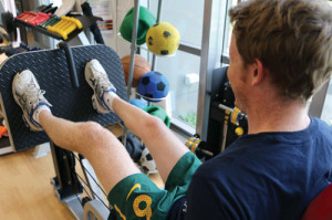
 Figure 1. A teenage patient with CP performs lower body strength training. (Image courtesy of the Cerebral Palsy Alliance.)


Figure 1. A teenage patient with CP performs lower body strength training. (Image courtesy of the Cerebral Palsy Alliance.)

But Sexton, along with the other experts that LER spoke with, doesn’t believe strength training (with diplegia or hemiplegia) is the ultimate panacea for gait and function issues in kids with CP. For strength training to be successful in this patient population, experts say, it needs to be part of a multifaceted rehabilitation program that takes into consideration more than the physical limitations imposed by the disease state.

“Children with CP are weak almost everywhere, so why not try to get them as strong as their able-bodied peers? Then get them to apply that strength and flexibility to whatever they want to do in their lives,” commented Jack Engsberg, PhD, director of the Human Performance Lab at Washington University in St. Louis. “I think the same principles for strength training that apply to people without disabilities apply to those with disabilities. But you have to consider what the disabilities are, and design a program based upon that.”

The goal of strength training in children with CP is not to produce the same results as body builders, stressed Christopher Joseph, MSPT, director of PT at the Kennedy Krieger Institute in Baltimore.

“We are looking to strengthen them within their abilities,” Joseph said.

Orthopedic surgeon Lance Silverman, MD, of Silverman Foot & Ankle in Edina, MN, said he is a believer in postsurgical PT in general, but cautioned, “Strength training by itself is a mistake because it will only strengthen the dysfunction instead of properly correcting the problem.”

A sound strength-training regimen will correct dysfunctional movement and then work to improve functional movement, Silverman explained.

“It is the same process that would be used with a healthy population; it’s just more challenging to do correctly with CP patients,” he said.

Patient selection

Study results for strength training in children CP have been mixed.1-6 Authors of a 2012 meta-analysis7 concluded that, while some individuals benefit from progressive strength training, it’s unlikely to be the optimal therapy for all patients with CP.

Engsberg, who is also a professor of occupational therapy, neurosurgery, and orthopedics, suggested the studies that did not show a good result from strength training did not aim for enough of a strength increase.

“These kids are already at thirty percent in terms of strength versus able-bodied kids, so a ten percent increase isn’t going to really benefit them,” he said. “You want to show a dramatic change in the strength component—sixty percent or more—so you have to tailor the training accordingly.”

But the experts agreed with the meta-analysis authors that patient selection is key. For example, kids with a Gross Motor Function Classification System (GMCFS) score of IV or V—in which independent mobility is either very limited or nonexistent—may not be good candidates for strength training.

“If I have a patient who I do not expect to walk after surgery, then I’m less likely to say that strength training is worthwhile,” Silverman said. “If I have a patient who I expect to have a productive gait after surgery, but is having a difficult time with balance and coordination, then I think strength training has value.”

The International Classification of Functioning, Health and Disability (ICF) has become a common tool for assessing disability, and ultimately, a child’s capacity for strengthening.

“The ICF considers the body structures and function aspect of a health condition/disability, the impact on activity, and the impact on participation,” explained Prue Golland, a consultant in physiotherapy at the Cerebral Palsy Alliance in Allambie Heights in New South Wales, Australia, in an email. “In simple terms, muscle weakness is considered to occur at the body structures level whilst walking is at the activity level. The literature8 suggests that interventions are generally effective at one level of the ICF only.”

Age and CP-related cognitive deficits are also considerations with regard to the child’s ability to follow directions.

Joseph explained that, at a very young age, most children with CP still learning effective motor patterns. If they struggle early on, that can lead to muscle weakness.

“When we start strengthening at an early age, we do it in a functional context. We’ll load them with a weight or put them in a context where they have to carry most of their body weight,” he said. “For instance, walking up steps. We may not start steps with a healthy eight-month-old because they can’t walk, but we may start steps in an eight-month-old CP kid because we want to strengthen the thighs and calves, and have the child learn the correct motor pattern.”

At the Cerebral Palsy Alliance, therapists reserve progressive resistance strength training for children older than 8 years; functional strength programs utilizing goal-directed therapy are used in younger children, Golland said.

Finally, Sexton said he does not advise strength training in children whose CP is complicated by severe cardiac abnormalities or bronchopulmonary dysplasia or in those with self-harming behavior.

Factors to consider

Strengthening programs are generally based on the guidelines from the American Academy of Pediatrics and the National Strength and Conditioning Association, Golland pointed out. But training approaches and protocols still need to be determined on a case-by-case basis.

At Kennedy Krieger, therapists use both the split treadmill and aquatherapy for strength training.

Figure 2. Tyler Sexton, MD, chats with a patient. (Photo by Bill Starling.)

Figure 2. Tyler Sexton, MD, chats with a patient. (Photo by Bill Starling.)

While motor activity is the primary aim of a split treadmill workout, it can offer some benefits for endurance and strengthening, Joseph said.

“Let’s say the child is hemiparetic on the right side. I can set her up so that the treadmill is going at the normal speed—about 1.5 to 2.2 miles per hour for an average child—on the left side. On the right, I can speed the treadmill up so that she has to concentrate more on that right leg,” he said.

A CP patient who has severe contraction and is unable to passively move her limbs may not be a candidate for cycling therapy, Sexton said, and would be better served by aquatic therapy or even resistance band training.

Sexton said he believes an effective strength-training regimen will incorporate antispastic medication, such as onabotulinumtoxinA (Botox) and baclofen. However, he emphasized that dosing of antispastics must be titrated properly to help control spasticity without limiting the patient’s ability to work their muscle groups and follow the training protocol.

Joseph concurred.

“Some kids use their spasticity and their muscle tone, even if it’s low, to function,” he said. “We don’t want to completely take away their spasticity and that muscle tone.”

AFOs can be a help or a hindrance, depending on the patient. Engsberg pointed out that, if a patient is wearing a rigid AFO but is trying to gain more ankle strength, then the AFO isn’t going to help with the latter if it prevents ankle motion.

“However, if a child is unable to walk without the AFO, then perhaps the goal with ankle strengthening might be to transition away from the rigid AFO to a flexible one,” he said.

And, even if an AFO does restrict ankle movement, the added stability may facilitate strengthening the muscles around the knee or hip, Golland said.

Joseph said that his group is getting away from orthoses in general and setting patients up with functional electrical stimulation (FES) units, in some cases while the patient is still wearing a postoperative cast.

“If we have concerns about the muscle getting weaker, we’ll cut a window in the cast and apply the FES, working within the parameters set by the physician,” Joseph said.

Time

One limitation of the published studies in this area is that the duration of strength training—generally around eight weeks—may not have been long enough for researchers to see visible functional improvements.

This is particularly true for patients who have recently undergone postoperative casting, Silverman said.

“It’s going to be several weeks after surgery before a CP patient is going to be able to recoordinate the body. You have to keep that timeline in mind before you determine when to begin strength training and how long it should be done,” he said.

Engsberg explained that a strength training protocol starts with learning proper technique, which in itself can take a couple of months.

“Then you start getting into the building of muscle mass,” he said. “So strength training programs that only go for four or six weeks are not really getting into the two important components that make strength training worthwhile.”

Rather than keeping a strict timeline for seeing results, the therapists at Kennedy Krieger follow the child’s progress in terms of functional gains, bearing in mind that, the younger the child, the more time progress will require.

“If we have a CP child who walks at age two, we’re going to work with them from twelve to sixteen months on strengthening in a functional context,” Joseph said. “As they get older, then we may be able to focus on a more traditional strengthening protocol because they can follow directions better.”

Sexton also emphasizes to his patients and their caregivers that they may not see the benefits of strength training in the short term.

“In the long run, they will have better mobility; they will have better range of motion. All of that is more likely to get them closer to their goals,” he said.

Goals and motivation

Sexton pointed out that many children with CP have the same hopes and dreams as kids without CP, which can be an important consideration in training.

“I wanted to be [basketball player] Shaquille O’Neal when I was younger,” he shared. “I remember the therapists would say to me, ‘Come on, Shaq, let’s do it,’ when I tried to get out of the wheelchair and walk. Was that the only thing that got me out of the wheelchair? No, but finding out what motivates a kid with CP and incorporating that into the strength training protocol is very important.”

For very young children who can’t communicate their goals, “you have to follow their lead and engage them as they are moving. Find out what seems to interest them, whether it’s walking the steps or being in the water,” Joseph said.

For some CP patients—especially those with GMFCS classification I or II—the goal may not be to improve function, but simply to maintain it, Golland said.

“Does function mean walking, getting up from the floor, or climbing stairs? Or does it refer to someone’s ability to reposition themselves in their wheelchair, or lean forward to assist a carer to reposition their clothing?”

Even small gains in strength may be empowering to a patient with CP.

“Let’s say a CP child is going through a calisthenics-based strength training program, but they’re only seeing a ten percent gain,” Sexton said. “From a clinical perspective, that may not be meaningful. But maybe that small gain allows that kid to feel like he can go out for a walk with his dad or do some other activity.”

Joseph noted that patient-centered quality of life is becoming essential for measuring the success of a training program.

“That kind of information about the child and his real-life situation can be much more useful [than more clinical measures] when I look at what is, and is not, working for the child,” he said.

Shalmali Pal is a freelance writer based in Tucson, AZ.

REFERENCES
  1. Engsberg JR, Ross SA, Collins DR. Increasing ankle strength to improve gait and function in children with cerebral palsy: a pilot study. Pediatr Phys Ther 2006;18(4):266-275.
  2. Unger M, Faure M, Frieg A. Strength training in adolescent learners with cerebral palsy: a randomized controlled trial. Clin Rehabil 2006;20(6):469-477.
  3. Eek MN, Tranberg R, Zügner R, et al. Muscle strength training to improve gait function in children with cerebral palsy. Dev Med Child Neurol 2008;50(10):759-764.
  4. Damiano DL, Arnold AS, Steele KM, Delp SL. Can strength training predictably improve gait kinematics? A pilot study on the effects of hip and knee extensor strengthening on lower-extremity alignment in cerebral palsy. Phys Ther 2010;90(2):269-279
  5. Scholtes VA, Becher JG, Comuth A, et al. Effectiveness of functional progressive resistance exercise strength training on muscle strength and mobility in children with cerebral palsy: a randomized controlled trial. Dev Med Child Neurol 2010;52(6):e107-e113.
  6. Scholtes VA, Becher JG, Janssen-Potten YJ, et al. Effectiveness of functional progressive resistance exercise training on walking ability in children with cerebral palsy: a randomized controlled trial. Res Dev Disabil 2012;33(1):181-188.
  7. Steele KM, Damiano DL, Eek MN, et al. Characteristics associated with improved knee extension after strength training for individuals with cerebral palsy and crouch gait. J Pediatr Rehabil Med 2012;5(2):99-106.
  8. Scholtes VA, Dallmeijer AJ, Rameckers EA, et al. Lower limb strength training in children with cerebral palsy–a randomized controlled trial protocol for functional strength training based on progressive resistance exercise principles. BMC Pediatr 2008;8:8:41.

New flatfoot data rekindle debate over role of obesity

$
0
0

2PEDS-obesity-iStock241274Australian researchers found no correlation between body mass index and prevalence of pediatric flatfoot, but used a different methodology than previous studies that reached an opposite conclusion. The conflicting results have revitalized the ongoing debate on this topic.
By Cary Groner

Over the years, researchers have claimed to discover significant correlations between childhood obesity and pediatric flatfoot. One well-known study by Pfeiffer et al, for example, examined 835 children aged 3 to 6 years and concluded that flatfoot prevalence was influenced by three factors: age (flatfoot was more common in younger children), gender (boys had a higher prevalence), and weight (42% of normal-weight kids had flat feet vs 51% and 62% of those who were overweight and obese, respectively).1 Other studies have reached similar conclusions.

Recent research has called such findings into question, however, and resurrected the controversy about which children should be treated. In a paper presented at the 2013 Australasian Podiatry Council Conference in Sydney last June2 and currently in press with the Journal of Foot and Ankle Research, Angela Evans, PhD, examined body mass index (BMI) and foot posture index (FPI) scores for 698 children and found no correlation between BMI and flatfoot prevalence. The work expanded on an earlier study reporting similar findings in 140 children.3

“These studies conflict with previous studies that found a correlation, even though intuitively that correlation makes sense,” acknowledged Evans, who has a private practice in Adelaide, Australia, and a research position at La Trobe University in Melbourne. “Part of the problem may be that most of those studies used a footprint-based method of assessing foot posture.”

Footprint measures raise an obvious question in the case of obese children: are the feet flatter or merely fatter? That is, when more adipose tissue is compressed under the child’s weight, does this merely create the impression of a flatter foot? The question has provoked significant debate—not only regarding the value of different assessment methods, but also in terms of its clinical implications.

The evidence

The evidence supporting the link between weight and flatfoot isn’t copious, but much of it seems plausible enough. For example, in a paper that appeared in 2006 in the journal Obesity, Australian researchers compared the feet of 19 overweight or obese preschool children with those of 19 controls matched for age, height, and sex. They concluded that there was no difference between groups in the thickness of the midfoot plantar fat pad, which was measured with ultrasound. But the heavier kids had a significantly lower plantar arch height, assessed via plantar footprints.4 Although footprints themselves may not offer a particularly robust evaluation tool, when the fat pad variable is accounted for, they seem more persuasive—or might, perhaps, in a larger sample size.

Another small study, this one from the US, reported similar findings in 2012, noting that obese children had less ankle dorsiflexion, resulting in longer foot contact time during stance, as well as significantly more flexible feet and greater arch drop.5

Some studies have been done in older children, which can complicate the comparison of results because their feet have had more time to develop. Nevertheless, a 2007 study of 200 Scottish children aged 9 to 12 years reported that foot length and width were greater, while navicular height was lower, in the heavier children.6 And a 2009 study from Spain evaluated foot arch types in 58 obese youths aged 9 to 16.5 years using both footprints and lateral weight-bearing radiographs, compared to the same number of normal-weight controls. Both measures found a lowering of the medial longitudinal arch in the obese children.7

A particularly interesting study was conducted in Spain and published in the European Journal of Pediatrics last year.8 Researchers used a 3D digital scanner to evaluate the foot morphology of 1032 schoolchildren aged 6 to 12 years, and reported that the arches of the obese children didn’t develop as fast as those of the normal-weight kids. A closer look at the reported data reveals odd contradictions, however. For example, the arch height of normal-weight children increased at an annual rate of 4.8%, whereas for obese children the rate was 3.7%. That fits the pattern; however, the rate for overweight kids, who were assessed separately from the obese ones, had a rate of 6.1%—higher than that of the normal-weight children.

Measurements

Clinicians and researchers can be forgiven for scratching their heads when faced with such data. Many now tend to draw conclusions tentatively, if at all. Angela Evans’s papers provide an example of how quickly things can shift, in fact.

In terms of her own research, she attributes the change of perspective to use of the FPI.

“The difference in our study that really stood out was the way we assessed foot posture,” she said. “We know that the FPI is fairly reliable; it has tested validity and is widely used clinically. So when we found these differences [vs previous research], we wondered what was going on. Our first study was small, but in the second one we got the same results in about seven hundred kids. There was not an association between increasing body mass and the rate of flat feet when measured by the FPI.”

Evans doubts the validity of footprint-based evaluations, and notes that even radiographic findings are problematic because there aren’t a lot of normative data to compare them with (not to mention the ethical issues that would be associated with subjecting schoolchildren to mass x-ray screening to gather those data).

But how about the FPI itself? Is it a valid assessment tool in obese children?

Anthony Redmond, MD, a researcher at the University of Leeds in the UK, who was largely responsible for developing and validating the FPI, told LER via email that it was originally designed as a measure for pediatric foot posture and was carefully validated in that role.9

“Re: childhood obesity, that is a different story altogether,” Redmond wrote. “[There are] problems with the use of footprint measures…and to some degree the same applies to the FPI and to any external measures of foot posture. Where the research question relates specifically to obesity, in my view the only valid measures of posture/alignment will be those that use internal imaging to visualise bone alignment directly.” Examples of such imaging modalities would include x-ray, magnetic resonance imaging, computed tomography, or radiostereometric analysis.

“I don’t think technically I absolutely agree,” Evans responded, when read this email. “The point is that the different measures have shown conflicting results. The FPI is widely used by clinicians, and I don’t think they have any sense of it being restricted regarding the incidence of childhood obesity.”

Clinical implications

Better clinical practice is, after all, the main point of sorting out such matters.

“Childhood obesity is terribly concerning from a wider health perspective,” Evans continued. “How much we need to worry about its relationship to foot posture is still undetermined, however, and I’m concerned that overdiagnosis may lead to unnecessary intervention.”

In the experience of Alan Ng, DPM, who practices with Advanced Orthopedics and Sports Medicine Specialists in Denver, CO, pediatric flatfoot has more to do with genetics than overweight.

“I lean toward agreeing with Evans on this issue, because in the kids I see, biomechanics are primarily based on genetics,” he said. “Later in life, if they maintain an elevated BMI, they may develop flatfoot secondary to overload and posterior tibial tendon dysfunction. But at that early stage, I don’t think childhood obesity really leads to flatfoot.”

Regardless of the patient’s weight, Ng decides whether to treat based on symptoms—and not just pain.

“If they come in complaining of tripping, or that they get too fatigued doing athletics at school, we assess them,” he said.

High on his list is checking for a secondary accessory ossicle or an os tibiale externum on the medial aspect of the navicular.

“They can have symptoms when they have a connection between that bone and the navicular, and the area gets irritated,” he explained.

Edwin Harris, DPM, associate professor of orthopedics and rehabilitation at Loyola University Medical Center in Maywood, IL, concurred with Ng about obesity and flatfoot.

“I tend to agree with the conclusions that increased BMI probably doesn’t correlate with pronated feet,” he said. “My patients include just as many skinny kids as obese kids, though it is harder to manage obese kids with orthotic devices.”

Harris pointed out that most of the literature relating BMI and flatfoot focus on arch height—which, as noted, is difficult to quantify clinically.

“There are no proven ways of measuring it unless you do it radiographically,” he said. “People say they’re going to measure the distance between the navicular and the floor, but I’m not sure how precise that measurement is, or whether I could duplicate it, due to all the variables.”

Harris also noted that researchers and clinicians are working without a particularly good definition of what constitutes flatfoot.

“It’s really a three-plane deformity, but most of the articles look at it only in the sagittal plane,” he explained. “That’s not really a good indicator of what’s going on.”

He, too, limits his treatment to those with symptoms, including pain in the arch.

“Pronated feet have a whole slew of morbidities other than obesity,” he said. “Some children are hypotonic or have other risk factors for muscle imbalance. But when they complain of pain, I listen to them and look at them carefully.”

Russell Volpe, DPM, who practices in Manhattan and is a professor of orthopedics and pediatrics at the New York College of Podiatric Medicine, said that the relationship between obesity and pediatric flatfoot is more complicated than simple cause and effect.

“Inactivity contributes to obesity, so we want to keep kids as active as possible,” he said. “From the podiatric perspective, if they have flatter, more pronated feet, and if that contributes to inactivity, then we have a vicious cycle that might lead to significant disability and worsening of obesity over time, which would be well worth avoiding.”

Asked whether only symptomatic children would likely curtain their activities, however, Volpe dissented.

“I’m not ready to concede that only symptomatic kids become less active,” he said. “There is such a thing as a presymptomatic child, and those of us treating children with flat feet need to be mindful of that. When we identify those, we need to do something to help them function better. Most of the time, when an older child or an adult comes to us with a mechanical foot problem, it’s been years in the making.”

Volpe considers BMI just one among a host of factors that help determine a clinical pathway.

“No kid is going to be in or out of the treatment group just because they’re heavy,” he said. “Obesity is interesting from a research, screening, and risk standpoint, but I’m not going to be the one saying, ‘Give all the fat kids orthotics.’ It’s just one more thing on my list of reasons when I decide to intervene or not.”

Volpe noted, moreover, that much of the discussion about obesity and flatfoot has turned on measurement.

“Angela points out that the footprint method is flawed; she hangs her hat on the FPI having normative value, and I’ll give her that,” he said. “But Tony Redmond now seems to be saying that the FPI is not the be-all and end-all, either. That Angela’s studies haven’t found a correlation may also be a measurement flaw, though in fairness to her, she acknowledges that in her conclusion. I’d be hard-pressed as a scientist and an academic to dismiss her work; I think it’s worthy of our attention. But I think we need to look further; there’s probably a bigger and better study to be done to see if we can settle this definitively.”

More studies

As it happens, recent research has shed light on which patients with flexible flatfoot are most likely to become symptomatic. In a study conducted at Rady Children’s Hospital in San Diego, researchers looked retrospectively at 135 patients, 45 of whom were asymptomatic. As assessed by standing anteroposterio and lateral radiographs, the authors concluded that lateral displacement of the navicular seemed to be related to symptom onset.10

“This may be an area we look at more carefully in the future, and it may start to simplify this whole debate,” said Evans.

In the meantime, however, she and her colleagues are about to embark on further research at La Trobe. The study will compare footprint measures, FPI, and the results of external 3D digitized foot scanning to evaluate their relative strengths and weaknesses.

“We need to clarify this discordance,” she said. “We’re hoping that this independent 3D imaging will teach us more about both of those measures. It’s quite exciting.”

Cary Groner is a freelance writer in the San Francisco Bay Area.

REFERENCES

1. Pfeiffer M, Kotz R, Ledl T, et al. Prevalence of flatfoot in preschool-aged children. Pediatrics 2006;118(2):634-639.

2. Evans A. The relationship between pediatric foot posture and body mass — do heavier kids really have flatter feet? J Foot Ankle Res 2013;6(Suppl 1):12.

3. Evans A. The paediatric flat foot and general anthropometry in 140 Australian schoolchildren aged 7-10 years. J Foot Ankle Res 2011;4(1):12.

4. Mickle KJ, Steele JR, Munro BJ. The feet of overweight and obese young children: are they flat or fat? Obesity 2006;14(11):1949-1953.

5. Shultz SP, Sitler MR, Tierney RT, et al. Consequences of pediatric obesity on the foot and ankle complex. J Am Podiatr Med Assoc 2012;102(1):5-12.

6. Morrison S, Dorward B, Watt G, Donaldson M. Anthropometric foot structure of prepubescent children with excessive versus normal body mass: a cross-sectional study. J Am Podiatr Med Assoc 2007;97(5):366-370.

7. Villarroya MA, Esquivel JM, Tomas C, et al. Assessment of the medial longitudinal arch in children and adolescents with obesity: footprints and radiographic study. Eur J Pediatr 2009;168(5):559-567.

8. Jimenez-Ormeno E, Aguado X, Delgado-Abellan L, et al. Foot morphology in normal-weight, overweight, and obese schoolchildren. Eur J Pediatr 2013;172(5):645-652.

9. Redmond A, Crane Y, Menz H. Normative values for the Foot Posture Index. J Foot Ankle Res 2008; 1(1):6.

10. Moraleda L, Mubarak S. Flexible flatfoot: differences in the relative alignment of each segment of the foot between symptomatic and asymptomatic patients. J Pediatr Orthop 2011;31(4):421-428.

In youth athletes, repair after meniscal injury poses challenge

$
0
0

2PEDSmeniscus-iStock169800Obesity, gender affect tear complexity
By P.K. Daniel

Adolescents and children suffer more complex meniscus injuries that are often less repairable than previously reported, according to a study published in the December 2013 issue of the American Journal of Sports Medicine (AJSM).

The study included 293 patients aged between 10 and 19 years who underwent arthroscopic meniscus surgery. Nearly all were active in sports, but factors contributing to greater complexity of the meniscal tears—which can adversely affect repairability—were obesity and male gender.

“Both obesity and boyhood carry an increased capacity for tear potential,” said study author Eric Edmonds, MD, who specializes in orthopedic surgery at Rady Children’s Specialists of San Diego in California.

Meniscal repair has demonstrated better long-term outcomes over partial meniscectomy in adults. A 2010 study showed no evidence of osteoarthritic advancement in 80.8% of patients a mean of 8.8 years after repair compared with 40% after meniscectomy.

“We recommend repair, if we can,” said Kelly Vanderhave, MD, who specializes in pediatric orthopedic surgery at Carolinas HealthCare System in Charlotte, NC. “Previous studies have shown that, if you take it out, you advance arthritis pretty quickly. If it’s at all repairable, I repair it.”

In the AJSM study, adolescent boys had a lower repair rate (41%) than adolescent girls (56%). Nearly one-third (32%) of the boys had complex tears, compared with just 20% of the girls studied, which also translated to lower repair rates among boys. Patients with complex tears also had a significantly higher body mass index than those with noncomplex tears (27.4 vs 25.1), though the study did not examine the relationship between body mass index and repair rates.

Meniscal injuries often present with other acute injuries such as anterior cruciate ligament (ACL) tears, chondral injuries, and tibial fractures. Although the correlation hasn’t been studied specifically, it’s likely that, just as pediatric anterior cruciate ligament (ACL) injury rates are on the rise, so too are pediatric meniscal injuries, Vanderhave said.

“They tend to go together,” Vanderhave said. “I think kids’ activity levels have increased overall. The number of sports injuries I see now is ten times what I saw ten years ago.”

Not all of the meniscal tears in the AJSM study were associated with ACL or other ligament injuries. The presence of ligament injury didn’t affect meniscal repair rates, said study coauthor Andrew T. Pennock, MD, who also specializes in orthopedic surgery at Rady Children’s Specialists of San Diego.

However, the authors did find that earlier treatment may increase the likelihood of repair in younger patients. Those treated within three months of injury were most likely to have repair, at a rate of 56%, compared with only 42% who were treated more than six months after injury. The repair rate for adolescents was even more dramatic, dropping from 58% at three months to 37% at six months.

Pennock noted a variety of reasons why surgeries are sometimes deferred, but delayed presentation led the list.

“Some are delayed presentation—the football player who didn’t want to come in midseason—some are patients who underwent a nonoperative course that failed, and others are delays in the workup, including a referral to the orthopedic surgeon, an MRI, and surgical booking or authorization,” he said.

Such delays increase the likelihood that a young, active patient will do further damage to the initial injury.

“Kids are very determined and resilient,” Edmonds said. “They often push through injuries because they don’t have the life experience to understand that you cannot just bounce back from all boo-boos. In that process, they continue to extend the injury past its original configuration. This can change from fixable to nonfixable.”

The surgical approach was typically determined by the type of the meniscal tear, in conjunction with the tear location, patient age, and chronicity of the tear. In the vast majority, surgeons did a partial meniscectomy if a repair was not possible.

“The reparability of the meniscus tear was determined by the surgeon at the time of surgery,” said Pennock. “In general, less complex tears in the red-white or red-red zone [at the outer edge of the meniscus] have the best healing potential and are the most likely to be repaired.”

P.K. Daniel is a freelance writer and editor based in San Diego, CA.

Sources:

Shieh A, Bastrom T, Roocroft J, et al. Meniscus tear patterns in relation to skeletal immaturity: children versus adolescents. Am J Sports Med 2013;41(12): 2779-2783.

Stein T, Mehling AP, Welsch F, et al. Long-term outcome after arthroscopic meniscal repair versus arthroscopic partial meniscectomy for traumatic meniscal tears. Am J Sports Med 2010;38(8):1542-1548.

Ankle weights improve walking in children with Down syndrome

$
0
0
2PEDSdown-DSC00843v2

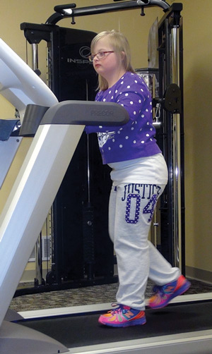
Photo courtesy of SureStep

Load may help engage plantar flexors

By Larry Hand

Adding an ankle load during treadmill walking significantly improves gait kinetics in children with Down syndrome (DS), possibly due to greater contributions from the ankle plantar flexors, according to a study from Georgia State University in Atlanta.

Jianhua Wu, PhD, assistant professor of kinesiology and health at Georgia State and coauthor Toyin Ajisafe, a graduate student, investigated effects of walking speed and external ankle load on the kinetic patterns of treadmill walking in preadolescents with DS and a comparable group of typically developing children.

Ten children with DS and 10 typically developing children (eight boys in each group; average age 9 years) participated in treadmill tests performed at two speeds (75% and 100% of preferred walking speed) and two ankle-load conditions (no ankle load or with ankle loads equal to 2% of the participant’s body weight). The added ankle load, applied using 1.25-lb weights from a sporting goods store, was intended to create a 39% increase in the moment of inertia for each leg at the hip joint.

The researchers instructed children to walk on the treadmill without placing their hands on the handrails, and provided verbal encouragement to the children with DS as needed during the tests. The researchers used a seven-camera motion capture system to collect kinetic and kinematic data, but, for this study, analyzed only the kinetic data.

Compared with the typically developing children, the children with DS had a shorter duration of propulsion during push off, a smaller second ground reaction force (GRF) peak and vertical propulsive impulse, a higher loading rate, and a lower unloading rate. The magnitude of the second GRF peak in the children with DS was smaller than body weight, a characteristic that is associated with typically developing children at younger ages, who tend to use the hip extensor muscles during push off rather than the ankle plantar flexor muscles.

Walking at a faster speed helped the children with DS improve the duration of propulsion, vertical propulsive impulse, and unloading rate, but the second GRF peak actually decreased further. However, the added external ankle load helped increase the second GRF peak as well as the vertical propulsive impulse, suggesting that the weights helped the children engage the ankle plantar flexors even at faster speeds. The findings were published in January by Gait & Posture.

“Inclusion of external ankle load may be a promising approach to strengthening leg muscles and eliciting a more powerful push off in persons with DS,” the authors wrote.

“Children with DS had little experience walking on a treadmill before this study,” Wu said. “However, the majority of them successfully walked on a treadmill at two speeds, slow and fast. It was surprising to their parents that their child could walk on a treadmill without holding the handrails. I think, with careful administration and appropriate monitoring, we can use certain paradigms such as treadmill walking to expand the motor repertoire of children with DS.”

This could help children with DS on more than a physical level, Wu said.

“A higher level of motor capability will certainly get children with DS more engaged in physical activity and sports. This may benefit children with DS not only at the physical health level, but also at the psychological and mental levels,” he said.

Kathryn Martin, PT, DHS, professor and Doctor of Physical Therapy program director at the Krannert School of Physical Therapy at the University of Indianapolis in Indiana, said the study highlights one possible way to improve strength and increase physical activity in children with DS.

“In my opinion, we cannot correct hypotonia or ligamentous laxity, but we can improve strength. Improving strength should help minimize compensations. Adding ankle load is one way to improve strength,” Martin said.

However, she noted that the effective use of treadmill technology in this patient population may be challenging for some clinicians.

“Children with DS always have some degree of intellectual disability,” she said. “Motivating them to walk on a treadmill may be more challenging than for typically developing kids, as the task may just not be meaningful to a child with DS. Physical therapists have to find ways to make these kinds of activities fun in order to encourage a child with DS to participate.”

Larry Hand is a writer in Massachusetts.

Source:

Wu J, Ajisafe T. Kinetic patterns of treadmill walking in preadolescents with and without Down syndrome. Gait Posture 2014;39(1):241-246.


Ankle dorsiflexion patterns vary in Charcot-Marie-Tooth disease

$
0
0
Will-running-and-kicking

Photo courtesy of the CMTA

Data support case-by-case approach

By Larry Hand

Gait patterns can vary significantly among young patients with Charcot-Marie-Tooth disease (CMT), and pathomechanics can vary even between limbs in one child, according to a recent study published in Gait & Posture.

This underscores the need for lower extremity practitioners to evaluate each child’s gait individually rather than rely on a textbook definition of CMT, according to study author Sylvia Õunpuu, MSc, director of research at the Center for Motion Analysis at Connecticut Children’s Medical Center in Farmington.

Õunpuu and colleagues retrospectively analyzed 33 children and adolescents (aged 12 ± 4 years) with CMT who underwent motion analysis and clinical examination while being evaluated for orthopedic surgery between 1990 and 2011. They compared data on patients with CMT with data on an age-matched control group of typically developing children.

Delayed peak dorsiflexion in terminal stance was seen in 59 of 66 limbs, and in some cases was the only gait abnormality found. This suggests delayed peak dorsiflexion may be the first gait-related sign of CMT, representing a major finding, according to Õunpuu.

Although the authors considered patient data initially as a single group, the evidence led them to subsequently divide patients into three subgroups based on peak ankle dorsiflexion in terminal stance, which is affected by plantar flexor length, plantar flexor strength, and cavus foot posture. Nineteen patients (30 limbs) had typical peak ankle dorsiflexion, eight patients (13 limbs) had less than typical dorsiflexion, and 14 patients (23 limbs) had excessive dorsiflexion. The findings were published in the September 2013 issue of Gait & Posture.

“One of the primary messages from this paper is that the orthopedic textbooks generally describe CMT as a certain set of conditions and, therefore, a certain set of treatments,” Õunpuu said. “CMT presents in many different ways and the degree of severity differs from patient to patient; therefore, treatment differs from patient to patient.”

In addition, gait pathomechanics may be asymmetrical in patients with CMT, she added, with some patients having one limb in the typical peak ankle dorsiflexion subgroup and the other limb in a different subgroup.

“Understanding that, and understanding maximum ankle dorsiflexion during gait and the extent of cavus deformity and plantar flexor strength and how these findings interact in each patient is really important for making an appropriate decision around treatment,” she said.

Ankle foot orthoses (AFOs) and other interventions can be used to address cavus foot presentation, excessive ankle dorsiflexion due to plantar flexor weakness, and plantar flexion contracture that results in limited dorsiflexion. However, clinicians need to be aware that overcorrection can create a new set of gait-related issues, the authors noted, and specified that surgical lengthening of the plantar flexors is not recommended.

“Two important goals an AFO should achieve are, first, to limit excessive dorsiflexion and allow weight bearing on the distal portion of the foot to provide more stability, and, second, to control cavovarus foot presentation to prevent plantar flexion contraction,” said Sean McKale, CO, LO, practice manager of Midwest Orthotic and Technology Center in Chicago, and a member of the Charcot-Marie-Tooth Association (CMTA) advisory board.

“This may be why we generally have had more success with ground reaction style and carbon fiber AFOs, and working with manufacturers to customize a stiffness profile for a particular individual. Additionally, using casting techniques and extrinsic lateral forefoot posting to maintain neutral coronal foot position has always been important to improve balance and distribute weight more evenly across the forefoot,” he said.

Practitioners are often better able to address gait-related CMT issues when treating patients as children than as adults, said David Misener, CPO, of Clinical Prosthetics and Orthotics in Albany, NY, who is also on the CMTA advisory board.

“Typically you’re able to push children a little bit harder,” Misener said. “Their bodies are little bit more elastic than adults’ and you can be a little bit more aggressive. Typically there are fewer psychosocial issues for children, and they are more willing to wear a device.”

Larry Hand is a writer in Massachusetts.

Source:

Õunpuu S, Garibay E, Solomito M, et al. A comprehensive evaluation of the variation in ankle function during gait in children and youth with Charcot-Marie-Tooth disease. Gait Posture 2013;38(4):900-906.

Ponseti method surpasses surgery for long-term clubfoot outcomes

$
0
0
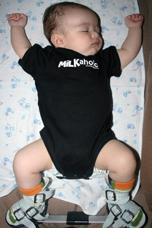
Milkaholic-2

Photo courtesy of MD Orthopaedics

But both groups fall short of controls

By Samantha Rosenblum

Children treated for clubfoot with the Ponseti method have less pain and fewer gait impairments as adults than those treated with surgery, according to research from Shriners Hospital for Children in Chicago.

In 42 adults aged between 19 and 35 years who were treated for clubfoot as infants (24 surgically, 18 using the Ponseti method) between 1983 and 1987, researchers compared the long-term results of each procedure in terms of foot function, foot biomechanics, and quality of life; they also compared the clubfoot patients to a control group of individuals with no history of clubfoot.

Both treatment groups had strength and motion deficits compared with the control group, but the group treated with the Ponseti method had better outcomes than the surgical group in many areas.

For example, individuals in the Ponseti group demonstrated significantly greater ankle plantar flexion range of motion (ROM), greater ankle plantar flexor and evertor strength, and a lower incidence of osteoarthritis in the ankle and foot. Additionally, the researchers found significantly higher pain levels in the surgical group than in the Ponseti group, as well as significantly reduced gait speed and stride length and more time spent in double support.

Overall, the study supports efforts to correct clubfoot with the Ponseti method, which minimizes surgery to joints to promote ROM and strength—essential elements of adult functioning in the years after treatment. The findings were epublished in November 2013 Clinical Orthopedics and Related Research.

LER-Resource-Guide-Products-clubfoot

Children in the Ponseti group began receiving treatment an average of 12.4 days after birth. The Ponseti method, developed by Ignacio Ponseti, MD, consists of manipulation of the foot and casting. Casts are changed weekly with gradually increasing correction and, at the final stage (after about four weeks), surgeons do a heel cord tenotomy to achieve a plantigrade position of the ankle and foot, according to Peter Smith, MD, principal investigator of the study and an orthopedic surgeon at Shriners Hospital for Children in Chicago. Patients then underwent abduction bracing with a Denis-Browne bar and straight laced shoes, which they wore full time for two months, followed by nighttime use for four years.

Children in the surgical group had been treated with conventional casting and had an inadequate response or were approaching age 18 months (the cut-off age for corrective surgery), and subsequently underwent surgery, a comprehensive clubfoot release with a Cincinnati incision.

Despite the growing popularity and positive outcomes associated with the Ponseti method, there are some situations in which surgery might still be used. According to John Herzenberg, MD, director of Pediatric Orthopedics at Sinai Hospital and director of the International Center for Limb Lengthening, both in Baltimore, MD, parents must play a large role in treatment for the Ponseti method to work effectively.

“Less parent participation and compliance is needed for surgery than the Ponseti approach,” Herzenberg said. “The Ponseti approach consists of three months of casting by the physician and then four years of bracing, which has to be supervised daily by the parents. Some parents are not able to maintain this protocol, for a variety of reasons.”

Herzenberg cited social and economic barriers as issues that can adversely affect compliance.

The study’s limitations may have had substantial effects on the data collected. Most importantly, there exists a strong possibility of selection bias, as all patients in the study who underwent Ponseti method therapy received that treatment at the University of Iowa Hospital and Clinics in Iowa City, where surgeons have used the Ponseti method for decades. All individuals in the surgery group were treated at the Shriners Hospital for Children in Chicago, where surgery was the standard of care at the time for patients in whom conventional casting failed. The Ponseti method is now the standard clubfoot treatment at Shriners.

Clubfoot patients treated in more recent years have likely benefited from advances in treatment protocols, Smith said.

“We are always trying to speed correction and prevent recurrence through better casting and bracing, and it is definitely a field that is improving,” he said. “We think the outcomes are a bit better, particularly in achieving initial correction of the foot into an abducted position to prevent recurrence and meticulous detail to bracing. But there is still an intrinsic nature of clubfoot that we don’t understand that seems to result in diminished strength and motion to a varying degree—even with the best current techniques.”

Samantha Rosenblum is a journalism student at Northwestern University in Evanston, IL.

Source:

Smith PA, Kuo KN, Graf AN, et al. Long-term results of comprehensive clubfoot release versus the Ponseti method: Which is better? Clin Orthop Relat Res 2013 Nov 19. [Epub ahead of print]

Juvenile hallux valgus: Shoe fit and other considerations

$
0
0

5PEDS-JHV-iStock26079472Austrian research suggests too-short shoes may contribute to the development of bunions in children, and genetics also appear to play a role. Most clinicians try to avoid surgery in young patients, instead turning to conservative strategies such as foot orthoses and night splints.

By Christina Hall Nettles

Two Austrian studies suggest that juvenile hallux valgus (JHV) is far more prevalent in young children than previously recognized and that the culprit is too-short shoes. But will this relationship apply to children in other countries?

Christian Klein, MD, Elisabeth Groll-Knapp, PhD, Michael Kundi, PhD, and Wieland Kinz, PhD, of the Institute for Environmental Health, Center for Public Health, at the Medical University of Vienna, recorded the hallux angle and length of 1579 individual feet among preschool children aged 3 to 6.5 years.1 Fewer than 25% of the children’s feet exhibited a straight position of the great toe, with the majority displaying lateral deviations at different degrees. The “great toe varied between a straight position and a hallux angle of up to 19°,”1 according to the researchers, who stated there were no known evidence-based normal values and ranges of hallux angle for “children of such a young age, either based on radiographic measurements” or on data using their external method.

Investigators stratified the study sample’s 858 participants by gender, geographic location (urban vs rural), and Austrian province and performed upright 3D measurement of the children’s feet using an external method based on footprints (to avoid exposing healthy children to x-rays). They then randomly selected days to measure the inside length of the children’s indoor and outdoor shoes and compared those measurements with the length of the feet. Nearly 90% of the participants’ indoor shoe sizes were too short, and nearly 70% of the children wore outdoor shoes of insufficient length.1

LER-Resource-Guide-Products-ped-footwear

“We showed that there is a significant relationship between too-short shoes and the hallux angle; the shorter the shoe, the higher the value of the hallux angle. Although currently we are not able to tell which dosage is harmful, for example, four hours per day during two months leading to an angle of fifteen degrees,” Kinz said.

The Austrian Federal Ministry of Health commissioned the same group of researchers to analyze children’s perceptions and parents’ awareness of properly fitted shoes compared with investigators’ measurements.2 They recorded data from more than 496 children aged 6 to 10 years and looked at approximately 1000 pairs of outdoor shoes, slippers, and house shoes. Children were asked to wear one shoe that was several sizes too small, and one shoe that was fitted correctly. Interviewers asked test subjects to walk in both shoes and then questioned them about the experience. They found the participants could not feel a clear difference in fit, and many children described the shoe that was markedly shorter than the foot itself as a good fit.2

“During our research projects and numerous ‘Measuring Days for Kids’ Feet’ held in Germany, Switzerland, and Liechtenstein, we were surprised to see how many children were wearing shoes that were much too short; up to five sizes too small. Oddly, all of these children were entirely convinced that their shoes fit perfectly,” Kinz said.

Shoe sizes increase about a half size every four months in children aged 3 to 5 years, with boys’ feet typically presenting one size longer and one size wider than girls’ feet at the same age.3

“We advise children’s feet need twelve to seventeen millimeters [about one half to three quarters of an inch] of extra space in their shoes. One should measure the length of the feet and also the inner length of the shoes using a homemade cardboard template or a tool such as the plus12,4 a measuring tool we developed for parents,” he added.

The team acknowledged the validity of studies that show heredity, pes planus, metatarsus primus varus, first metatarsal length, and hypermobility of the metatarsocuneiform joint are significant factors in understanding the etiology of JHV. However, they believed no studies of extrinsic factors had investigated a significant relationship between wearing shoes of insufficient length and lateral deviation of the great toe.

Intrinsic vs extrinsic factors

Hallux valgus is the most commonly reported forefoot deformity in adults, yet a 2010 systematic review of papers reporting results of nearly 497,000 participants found its actual prevalence difficult to estimate consistently, and concluded only that the condition is common and is diagnosed more often in women and with increasing age.5

In juvenile imaging studies, normal hallux valgus angles range below 16°, and a normal intrametatarsal angle must not exceed 9°,6 but singular measurements may not be enough to make an accurate diagnosis. Juvenile hallux valgus is often bilateral, and an “increased distal metatarsal articular angle may be the defining characteristic of JHV,” according to a 1995 study7 of 60 feet that were treated with a variety of surgical corrections individualized to their specific deformities. Michael J. Coughlin, MD, current clinical professor of orthopedic surgery at the University of California San Francisco and past president of both the American Orthopaedic Foot and Ankle Society and the International Federation of Foot and Ankle Societies, led the investigation.

Coughlin acknowledged the importance of properly fitting shoes, but questioned if studies based in central Europe would apply with equal measure to children in other countries.

“Insufficient length is an interesting factor, but I think it plays a minor role in America, where children have multiple shoes and purchase shoe wear frequently,” Coughlin said. “In adolescent hallux valgus, I believe shoe wear plays a minor role in development and progression of deformity. We specifically asked our patients about shoe wear, and a small percentage felt this was a factor.”

Coughlin’s 1995 study was an 11-year retrospective review of 45 children (60 feet) with JHV. It found that constricting footwear was noted in only 24% of patients, but that nearly 75% of the study cohort inherited the deformity from their mothers.7 The latter finding was supported by a 2010 study of bilateral scarf osteotomy performed in adolescents with JHV, which found 100% maternal inheritance of hallux valgus deformity.8

Coughlin suggested that medical providers evaluating suspected JHV take a family history to determine the incidence of bunion deformities in parents, siblings, and close relatives, and take a closer look at patients with a hallux valgus angle greater than 25° aged between 12 and 14 years.

“Most children with JHV experience onset at ages seven to ten years or earlier. The age of onset is associated with increased severity of deformity. Family history and early onset make a difference in the severity—and often the progression—of deformity,” he told LER.

Coughlin’s research found no increase of pes planus in patients with JHV. Foot pronation may predispose hallux valgus in certain patients, such as those with ligamentous laxity, but research has not quantitatively demonstrated this predisposition.9

In 1993, a critical literature review noted JHV is often inherited and is more common in girls and in shoe-wearing populations, yet studies searching for the etiology of JHV had failed to prove the deformity was a product of the shape and length of the first metatarsal.10 More recent studies have continued exploring the associative or causative relationship of the first metatarsal in JHV.11

Management strategies

Norman Otsuka, MD, Joseph Milgram professor of orthopedic surgery at NYU Langone Medical Center in New York City, and orthopedic surgery resident Rachel J. Shakked, MD, typically see adolescent hallux valgus in female patients with a positive family history.

“Standing radiographs can calculate the intrametatarsal angle to confirm diagnosis, but the main factors in deciding whether to refer a patient for surgical consultation are the presence of pain and the need to significantly modify activities,” Shakked said.

“Diagnosing JHV in younger patients, male and female, is approached similarly—determining whether the foot deformity is painful and affecting activities,” she continued. “Conservative management may be advised unless pain, functional deficit, rapid progression, or other concerning signs are present. In younger patients, we advise nonoperative management when possible, as there is a high recurrence rate of hallux valgus after surgery if a patient is skeletally immature.12 Conservative treatment can include shoe modification to a wide toe box and night splints to abduct the great toe.”

In a 1992 study of 56 children with hallux valgus (aged one month to 16 years), night use of a thermoplastic splint along with exercises were associated with improved metatarsophalangeal joint angle, intermetatarsal angle, or both in about half the feet treated.13 Researchers reported no recurrences among those successful outcomes at follow-up, which ranged from two to six years.

Conservative interventions for hallux valgus may provide relief from symptoms, but may not reverse the deformity, which in some cases may be severe enough to dislocate the first metatarsophalangeal joint, according to Ward M. Glasoe, PhD, PT, ATC, assistant professor in the Physical Therapy Program at the University of Minnesota Medical School in Minneapolis.

Researchers have not been able to determine consistently which conservative interventions are appropriate in children who have not reached skeletal maturity. Glasoe cited a landmark study by Kilmartin et al14 indicating foot orthoses worsened the existing JHV angle in children aged 9 to 10 years, but he noted the study was limited by its use of rigid partial-contact foot orthoses designed to limit hindfoot pronation and by an unverifiable measure of patient compliance.

“More current evidence recognizes the benefits of treating hallux valgus with foot orthoses,” Glasoe wrote in Physical Therapy.15 “Orthoses that bolster the arch and orient the first metatarsal horizontally may work to contain the kinetics and kinematics of the first metatarsal to the sagittal plane.”

“Orthoses posted medially combined with strengthening exercise of the tibialis posterior, in my clinical experience, offer a chance to correct flattening of the arch and excess eversion of the hindfoot and possibly counteract the progression of hallux valgus,” Glasoe said.

Seattle podiatrist Lawrence Z. Huppin, DPM, of The Foot and Ankle Center of Washington, fits custom and customized foot orthoses for children and adolescents with JHV. He, too, looks closely at family history when determining how aggressively to treat young patients with hallux valgus.

“In adolescent patients, our primary goal is to allow the first ray to plantar flex. When we cast for orthoses, we plantar flex the first ray so that the resultant orthosis is more effective at preventing jamming of the first midtarsophalangeal joint. When writing the orthotic prescription, we use a minimum cast fill and several degrees of inversion in the positive cast, so the orthosis conforms closely to the arch in order to allow that first ray to plantar flex. If the rearfoot is everted, we prescribe a device with a deeper heel cup and a medial heel skive to prevent further eversion,” he told LER.

“In younger children, I am more likely to prescribe a prefabricated orthosis that offers these features [deep heel cup, medial skive, forefoot valgus correction, and a fairly high arch], because children grow out of them quickly and they can become prohibitively expensive. Adolescents’ custom orthotics are likely to last for several years.”

Pain is the most common reason for surgical intervention in JHV, with progression of deformity another major factor to consider, Coughlin reported.

“On the other hand, we sometimes wait until a patient is a bit older to perform surgery,” he said. “It is not imperative that growth lines be closed, but surgery must be carefully performed to avoid injury to the growth plate.”

Several surgical procedures have been shown to successfully correct JHV, but as Coughlin and Shakked noted, concerns remain about performing orthopedic surgery in skeletally immature patients.

Shakked clarified that postsurgical therapy depends on the type of surgery performed.

“Aggressive range-of-motion activities should be minimized if an osteotomy is done to allow the osteotomy site to heal, which usually takes about six to eight weeks,” she said. “The surgeon may apply a bandage with a toe spica tape splint to keep the soft tissues stretched on the lateral side of the first MTP joint. Stretching activities of these ligaments and strengthening the abductor hallucis can be helpful postoperatively. After the bone has healed, surgeons may recommend range-of-motion activities of the hallux to prevent stiffness and regain preoperative range of motion.”

Physical therapy after any mode of JHV surgery may require extra measures to protect the surgical site and the rehabilitation of posture and gait.

In an earlier LER article exploring physical therapy after bunion­ectomy in adults, Clarke D. Brown, PT, DPT, OCS, ATC, who is in private practice in Macedon, NY, and is president of the American Physical Therapy Association’s foot and ankle special interest group, noted that postsurgical physical therapy should extend well above the ankle, and protocols must be adapted for individual patients (see “Beyond bunionectomy: The role of physical therapy,” August 2010, page 18). He further advised unique factors to consider when evaluating children who have undergone osteotomy for JHV.

“Young adults can be impetuous and impatient. If their surgery restores weight-bearing activity or relieves pain, these patients may push weight bearing by standing or walking on the surgical side earlier than is appropriate,” Brown said.

“Great attention should be paid to making sure surgical sites are well protected, proximally and distally. Younger children will often retain gait and movement deviations learned during painful periods. Many will continue to needlessly limp or substitute during gait, because their motor patterns were chronically changed. It is often difficult to restore normal movement even when pain and mobility are corrected,” he cautioned.

Next steps

Further studies by Kinz’s team in Austria will explore the effects of intervention programs that seek to ensure correctly fitting shoes and assess whether hallux valgus is reversible after changing footwear habits.

Coughlin led a preoperative study of demographics, etiology, and radiographic findings in adults with moderate to severe hallux valgus deformities and found that 34% of patients implicated constricting shoes or occupation as causes of their bunions.16 However, nearly a quarter of participants had developed the deformity at age 20 years or younger. Family history, female gender, a long first metatarsal, and an oval or curved metatarsophalangeal joint articular surface were common findings.

Clearly, more research is needed to identify the role of poorly fitting footwear and its causative or associative function in the early development of JHV, but the persistent inability of children to distinguish poorly fitting from properly fitting shoes reported in the Austrian studies is a critical observation.

“These studies are a reminder that any of us who treat children’s feet should take the opportunity to educate parents about proper fit,” Huppin said. “Children’s feet are more pliable than adults’. While shoe length certainly is important, we also need to make sure children’s shoes are wide enough for their toes and are not applying external medial or lateral force.”

Christina Hall Nettles is a freelance writer based in Monroeville, AL.

REFERENCES

1. Klein C, Groll-Knapp E, Kundi M, Kinz W. Increased hallux angle in children and its association with insufficient length of footwear: a community based cross-sectional study. BMC Musculoskelet Disord 2009;17(10):159.

2. Forschungsprojekt “Kids: healthy feet—healthy life.” https://www.kinderfuesse.com/presseinfo/presseinfo2007_2_en.html. Published March 26, 2007. Accessed April 3, 2014.

3. Gould N, Moreland M, Trevino S, et al. Foot growth in children age one to five years. Foot Ankle 1990;10(4)211-213.

4. Plus12. Kinderfuesse.com website. https://www.kinderfuesse.com/english/plus12. Accessed April 3, 2014.

5. Nix S, Smith M, Vicenzino B. Prevalence of hallux valgus in the general population: a systematic review and meta-analysis. J Foot Ankle Res 2010;3:21.

6. Easley ME, Wiesel SW. In: Operative Techniques in Foot and Ankle Surgery. Philadelphia: Lippincott Williams & Wilkins; 2010:11-12.

7. Coughlin MD. Roger A. Mann Award. Juvenile hallux valgus: etiology and treatment. Foot Ankle Int 1995;16(11):682-697.

8. John S, Weil L Jr, Weil LS Sr, Chase K. Scarf osteotomy for the correction of adolescent hallux valgus. Foot Ankle Spec 2010;3(1):10-14.

9. Coughlin MJ, Saltzman CL, Anderson RB, ed. Hallux Valgus. In: Mann’s Surgery of the Foot and Ankle, 9th ed. Philadelphia: Saunders; 2014:169-170.

10. Kilmartin TE, Wallace WA. The etiology of hallux valgus: a critical review of the literature. The Foot 1993;3(4):157-167.

11. Vyas S, Conduah A, Vyas N, Otsuka NY. The role of the first metarsocuneiform joint in juvenile hallux valgus. J Pediatr Orthop B 2010;19(5):399-402.

12. Ball J, Sullivan JA. Treatment of the juvenile bunion by Mitchell osteotomy. Orthopedics 1985;8(10):1249-1252.

13. Groiso JA. Juvenile hallux valgus. A conservative approach to treatment. J Bone Joint Surg Am 1992:74(9):1367-1374.

14. Kilmartin TE, Barrington RL, Wallace WA. A controlled prospective trial of a foot orthosis for juvenile hallux valgus. J Bone Joint Surg Br 1994;76(2):210-214.

15. Glasoe WM, Nuckley DJ, Ludewig PM. Hallux valgus and the first metatarsal arch segment: a theoretical biomechanical perspective. Phys Ther 2010;90(1):110-120.

16. Coughlin MJ, Jones, CP. Hallux valgus: Demographics, etiology, and radiographic assessment. Foot Ankle Int 2007:28(7)759-777.

Late-breaking ACL data raise questions about use of landing-based screening

$
0
0

shutterstock_911573-lrBy Jordana Bieze Foster

A presenter from Norway gave IOC conference attendees a sneak peek at his group’s latest findings and generated considerable discussion by suggesting that many aspects of drop-landing mechanics are not predictive of anterior cruciate ligament (ACL) injury in elite female athletes.

Eirik Kristianslund, MD, a researcher at the Oslo Sports Trauma Research Center, presented the results not as part of a Free Communications session, but as part of a symposium on screening for ACL injury using drop-jump tasks.

Kristianslund and colleagues analyzed landing mechanics in 708 elite female handball and soccer athletes (mean age, 21 years) as they performed a drop jump from a 30-cm height, followed by a maximum-height jump after landing. During seven years of follow-up, 38 players sustained ACL injuries.

Knee valgus angle at initial contact, maximum knee adduction moment, peak vertical ground reaction force, and maximum knee flexion angle were not significantly associated with ACL injury risk. Of the variables measured, the only one that was significantly associated with ACL injury risk was medial knee motion, with an odds ratio of 1.35. However, Kristianslund noted that the average between-group difference in medial knee motion was only 5 mm, and questioned whether that measure would be clinically useful for screening.

Although the findings do not support drop-jump screening to identify high-risk individuals in this population of elite athletes, many of whom may have previously undergone neuromuscular training, Kristianslund cautioned that the results may not apply to other populations.

medi_LER_igli_DigitalAd_FINAL

Poor dynamic postural stability predicts risk of ankle injury in ball team athletes

$
0
0

iStock_000018439322_smBut exercises may not help those with CAI

By Jordana Bieze Foster

Poor dynamic ankle stability is predictive of injury risk in athletes, but exercises designed to improve dynamic balance may not be effective in athletes with chronic ankle instability, according to separate studies presented at the IOC conference in Monaco.

Investigators from the University Medical Centre Groningen in the Netherlands assessed dynamic postural stability and drop jump-landing technique in 42 athletes (mean age, 22.2 years) who played basketball, korfball, or volleyball at an elite level and tracked knee and ankle injury rates for one year. Dynamic postural stability index (DPSI) scores, with high scores indicating poor stability, were based on mediolateral, anterior-posterior, and vertical stability (time to stabilization) associated with single-leg jump landings onto a force plate in frontal, diagonal, and lateral directions. Drop-landing technique was assessed using the Landing Error Scoring System (LESS; higher score reflects poorer technique).

One year later, the Dutch researchers found that athletes who went on to suffer an ankle injury had significantly higher baseline DPSI scores for diagonal anterior-posterior, diagonal vertical, and lateral anterior-posterior components than those who had no ankle injuries. Athletes who went on to sustain a knee injury had higher baseline LESS scores than those with no knee injuries, but that difference was not statistically significant.

medi_LER_igli_DigitalAd_FINAL

Those findings suggest there may be an opportunity for intervention, such as balance exercises, to prevent ankle injuries in athletes with elevated DPSI scores. But a Belgian study presented at the IOC conference found that balance training did not significantly improve DPSI scores in recreational athletes with chronic ankle instability (CAI).

In 64 recreational athletes, 33 of whom had chronic ankle instability, researchers from Ghent University assessed DPSI before and after a home-based progressive balance exercise program. Athletes performed exercises three times per week for eight weeks. Progressions included adding arm movements, going from eyes open to eyes closed, and decreasing the stability of the exercise surface.

Baseline DPSI scores confirmed that dynamic postural stability was significantly poorer for the athletes with CAI than the controls, as were self-reported functional scores on the Foot and Ankle Disability Index (FADI). After the intervention, the FADI scores had improved significantly in the CAI patients, but the DPSI scores had not.

It may be that the balance exercises were not specific enough to improve dynamic measures, or that the DPSI was not sensitive enough to detect more subtle degrees of improvement, according to Roel De Ridder, a doctoral student in the Department of Physical Therapy and Motor Rehabilitation at the university, who presented his group’s findings in Monaco.

“I think athletes should do balance training, but to improve postural control we have to potentially think about incorporating other exercises or training techniques,” De Ridder said.

Another study from the same Belgian group may shed some light on the landing kinematics underlying the postural control deficits seen in athletes with CAI. In 96 recreational athletes, De Ridder and colleagues found that those with CAI had a stiffer landing technique during vertical drop jumps and side jumps than controls or “copers” who had a history of ankle sprain but did not have ongoing instability. A multisegmented foot model further distinguished CAI patients from copers, who demonstrated less plantar flexion and dorsiflexion range of motion in the lateral and medial forefoot.

Sources:

Van der Does H, Brink M, Lemmink K. A one year prospective study on ankle stability and landing technique: The occurrence of ankle and knee injuries in elite ball team athletes. Br J Sports Med 2014;48(7): 586.

De Ridder R, Willems T, Vanrenterghem J, Roosen P. Effect of balance training on dynamic postural control in subjects with chronic ankle instability. Br J Sports Med 2014;48(7):584.

De Ridder R, Willems T, Vanrenterghem J, et al. Multi-segmented foot landing kinematics in subjects with chronic ankle instability. Br J Sports Med 2014; 48(7):584.

UTC monitoring of patellar tendon load keeps Australian footballers in the game

$
0
0

shutterstock_107619803lrBy Jordana Bieze Foster

Using ultrasound tissue characterization (UTC) to monitor tendon load in Australian Football League players has led to no games missed due to patellar tendinopathy in four years, according to Sam Rosengarten, a sports physiotherapist at Recover Sports Medicine in Melbourne who has worked most recently with the Carlton Football Club.

Rosengarten discussed his experience with UTC as part of a symposium on patellar tendinopathy at the IOC conference in Monaco. The technology synthesizes 3D ultrasound data into a color-coded image that reflects tissue integrity.

This allows team practitioners to carefully monitor athletes to prevent overloading of tissues, which become apparent on a UTC scan long before symptoms develop. It also helps to avoid the opposite problem of undertraining. The typical AFL team averages one case of patellar tendinopathy per season, which leads to an average of nearly three games missed—something Rosengarten says UTC monitoring can help avoid.

“With vigilant monitoring with UTC we can make good decisions about how much to load and how often. Our ability to intervene at the reactive stage and stop them from progres­sing to the degenerative stage is an amazing opportunity,” Rosengarten said. “Once they get to the degenerative state there’s almost no chance of getting back to a normal-looking tendon.”

medi_LER_igli_DigitalAd_FINAL

Support builds for eccentric strengthening to prevent hamstring strains in soccer

$
0
0

iStock_000022097896_smNordic exercise could be used for screening

By Jordana Bieze Foster

Research is accumulating in support of eccentric strengthening exercises to prevent hamstring injury in soccer players, and possibly to screen for athletes at risk, according to several studies presented at the IOC conference in Monaco.

Investigators from the Netherlands and the US both found that male soccer players who performed eccentric strengthening exercises were significantly less likely to sustain hamstring injuries than those who did more conventional training.

In the Dutch study, 41 teams of male amateur soccer players were randomized to 13 weeks of regular soccer training or training that included 25 sessions of Nordic hamstring exercises (also known as Nordic hamstring lowers). The results presented in Monaco included analysis of 16 teams in the intervention group and 19 teams in the control group.

medi_LER_igli_DigitalAd_FINAL

In the nine months following the intervention, there were 19 hamstring injuries in the control group and six in the intervention group, a statistically significant difference that translates to a relative risk of 4.267 for the controls. Number of days missed per injury, however, did not differ significantly between groups, according to Nick van der Horst, MSc, a researcher in the department of rehabilitation at the University Medical Center Utrecht, who presented the findings.

In the US, a study of collegiate soccer players found that a significant reduction in hamstring injury risk appears to be a benefit of using the FIFA 11+ warm-up program, which includes eccentric hamstring strengthening.

Researchers from the Santa Monica Sports Medicine Foundation in California analyzed male soccer players from 61 college teams, 27 of which implemented the FIFA 11+ program three times per week for one competitive season. They found that the rate of hamstring injuries was 74% lower in the intervention group than in the control group, according to Holly Silvers, MPT, director of research for the foundation, who presented the findings in Monaco. (In addition, the rate of anterior cruciate ligament injuries was 58% lower in the intervention group.)

“We’ve got the science. We know the program is working. Now we just have to get people to utilize it,” Silvers said.

The Nordic hamstring exercise may also be useful as a screening tool to identify athletes at increased risk for hamstring injury, according to research from Queensland University of Technology in Brisbane, Australia. Of 210 elite Australian football players, those with low levels of eccentric strength during the Nordic hamstring exercises at the start of the preseason were significantly more likely to go on to sustain a hamstring injury.

But increasing that strength may be easier said than done, particularly in athletes with a history of hamstring injury, which in itself is a risk factor for future injury. A second study from the Queensland group found that Australian football players with a history of hamstring injury demonstrated significantly less improvement in knee flexor strength than uninjured players following 16 to 18 weeks of training that included Nordic hamstring exercises.

Interestingly, the limited improvement in the players with previous hamstring injury was observed in both limbs, not just the previously injured limb, according to Anthony Shield, PhD, a senior lecturer in the School of Exercise and Nutrition Sciences at the university, who presented the findings in Monaco.

Sources:

Van der Horst N, Wouter Smits D, Petersen J, et al. The preventive effect of the Nordic hamstring exercise on hamstring injuries in amateur soccer players: A randomized controlled trial. Br J Sports Med 2014;48(7): 609-610.

Silvers H, Mandelbaum B, Bizzini M, Dvorak J. The efficacy of the FIFA 11+ program in the collegiate male soccer player (USA). Br J Sports Med 2014;48(7):662.

Opar D, Williams M, Timmins R, et al. Eccentric hamstring strength during the Nordic hamstring exercises is a risk factor for hamstring strain injury in elite Australian football: A prospective cohort study. Br J Sports Med 2014;48(7):647-648.

Shield A. Previous hamstring injury reduces pre-season improvements in eccentric strength in elite Australian football players. Br J Sports Med 2014; 48(7):660.


Education before ACL reconstruction reduces rates of reinjury in first year

$
0
0

5IOC-Running-iStock4393486lrBy Jordana Bieze Foster

Two hours of patient education before anterior cruciate ligament (ACL) reconstruction can significantly reduce rates of reinjury in the first year after surgery, according to research from Funabashi Orthopedic Hospital in Japan.

Investigators analyzed 252 ACL reconstruction patients who voluntarily chose to attend a two-hour lecture prior to undergoing surgery and 200 patients who chose not to attend. The same surgical technique was used in all patients, and patient activity levels were similar between the two groups.

The lecture, which included a slide presentation and patient handouts, has been offered to ACL reconstruction patients since 2003. It covers ACL anatomy, a description of the operative procedure, rehabilitation, and risk factors for reinjury. Patients who attended the lecture were told to expect about 10 months of rehabilitation before being able to return to sports participation.

At a mean follow-up of 20.9 months, the number of subsequent ACL injuries did not differ significantly between the two groups. However, the reinjury rate in the first year after surgery was significantly lower in the patient education group than the control group, according to Kenji Sato, PT, a researcher at the hospital who presented the group’s findings at the IOC conference in Monaco.

Source:

Sato K, Tsuchiya A, Kanisawa I, et al. The effect of educational lecture on reducing reinjury after anterior cruciate ligament reconstruction. Br J Sports Med 2014;48(7):658.

medi_LER_igli_DigitalAd_FINAL

Research downplays role of shoe design elements in maintaining runners’ health

$
0
0

5IOC-Running-iStock31482254lr‘Comfort filter’ may reduce injury risk

By Jordana Bieze Foster

Runners today aren’t like the runners of the 1970s. They’re far less likely to be male, thin, or dedicated to the sport. And yet, the medical literature suggests that running injury rates are essentially unchanged.

How much of this can be attributed to footwear design? Longtime biomechanist Benno Nigg, Drscnat, Drhcmult, who gave a keynote on the topic at the IOC conference in Monaco, isn’t sure.

“The effects of shoes on running and running injuries are not well understood,” said Nigg, a professor of biomechanics and co-director of the Human Performance Laboratory at the University of Calgary in Alberta, Canada, who is set to retire at the end of this summer.

The one footwear characteristic Nigg does believe affects injury risk in runners is comfort. This is based on the findings of a 2001 study in which his group found that injury rates in military personnel were significantly lower in those who wore inserts they selected based solely on comfort than in those who wore no inserts. The selected inserts varied widely with regard to component materials.

medi_LER_igli_DigitalAd_FINAL

“We all have different optimal shoes, but we all also have a comfort filter, and that reduces injury risk,” Nigg said. “That’s why running injuries haven’t changed—because you always run in shoes that you like.”

Several studies presented at the IOC conference tend to support the theory that specific footwear design characteristics do not influence injury rates.

In 247 runners randomized to use a shoe with a hard midsole or one with a soft midsole for five months, researchers from the Public Research Centre for Health in Luxembourg found that the risk of running-related injury was similar between groups.

And in a study of 1696 novice runners, investigators from the University of Groningen in the Netherlands found that the incidence of running-related injuries over six weeks was not significantly related to the age of running shoes worn.

In a third study, the Luxembourg researchers found that nine-month follow-up data confirmed earlier five-month results, suggesting that runners who switched between different pairs of shoes had a lower risk of running-related injury than those who predominantly used only one pair of shoes. The nine-month hazard ratio for multiple shoe use was .539, even lower than the hazard ratio of .614 seen at five months (see “Switch trials: Shoe use, striking affect risk in runners”).

Multiple-shoe users were about three times more likely to switch between different brands of shoes than between different models of the same brand or different versions of the same model—which again appears to support the idea that particular shoe characteristics do not influence injury risk. Rather, it may be that sticking with any type of shoe for too long, regardless of its structural features, is detrimental, said Laurent Malisoux, PhD, a researcher at the Luxembourg center’s Sports Medicine Research Laboratory, who presented the findings in Monaco.

“We cannot conclude from our findings any causal relationship, but I think that multiple shoe use is protective against injury,” Malisoux said. “It could induce a variation in external or internal forces, and we know that overuse injury is caused by repetitive microtrauma.”

Sources:

Mundermann A, Stefanyshyn DJ, Nigg BM. Relationship between footwear comfort of shoe inserts and anthropometric and sensory factors. Med Sci Sports Exerc 2001;33(11):1939-1945.

Theisen D, Malisoux L, Delattre N, et al. Does running shoe midsole hardness influence running-related injuries? Results from a double blind randomized controlled trial. Br J Sports Med 2014;48(7):664.

Kluitenberg B, van der Worp H. Risk factors for running related injuries in novice runners participating in a 6-week running program. Br J Sports Med 2014;48(7): 620-621.

Malisoux L, Urhausen A, Theisen D. Impact of training characteristics on running-related injuries in recreational runners. Br J Sports Med 2014;48(7):631-632.

Malisoux L, Ramesh J, Mann R, et al. Can parallel use of different running shoes decrease running-related injury risk? Scand J Med Sci Sports 2013 Nov 28. [Epub ahead of print]

Specialization, weekly training loads contribute to risk in youth athletes

$
0
0

5IOC-implementation-shutterstock_104766692lrBy Jordana Bieze Foster

Sports specialization and high weekly training volumes are associated with increased risk of injury in youth athletes, according to research from Loyola University in Chicago.

Investigators compared 837 injured athletes aged between 7 and 18 years recruited from sports medicine clinics and 353 uninjured youth athletes presenting for sports physicals at primary care clinics.

Injured athletes averaged significantly more hours per week playing sports than uninjured athletes (11.3 vs 9.4 hours, respectively). Increased injury risk was associated with playing sports for more hours per week than the child’s age and playing organized sports for twice the number of hours per week as free play.

Highly specialized athletes, in particular those who had quit all other sports to focus on just one, were most likely to have a serious overuse injury—even after adjusting for number of hours played per week. Privately insured athletes were more likely to specialize and also more likely to report a serious overuse injury.

Annual growth rates, however, did not differ significantly between the injured and uninjured groups.

“We do think there is an independent risk of sport-specific training for injury and for serious overuse injury,” said Neeru Jayanthi, MD, an associate professor of family medicine at the university, who presented the findings at the IOC conference.

Source:

Jayanthi N, Dugas L, Fischer D, et al. Risks of intense, specialized training and growth for injury in young athletes: A clinical evaluation. Br J Sports Med 2014;48(7):611.

medi_LER_igli_DigitalAd_FINAL

Studies explore implementation options for sports injury prevention programs

$
0
0

5IOC-implementation-iStock16877226lrClosing the gap between lab and field

By Jordana Bieze Foster

Even if an injury prevention program is effective under controlled study conditions, that effectiveness doesn’t always translate to the real world. That’s why some researchers are now working to identify the most effective strategies for implementing prevention programs and the most common barriers to implementation.

One challenge in implementing a neuromuscular training program is finding qualified people to train the athletes. Coaches are often too busy or lack the physical fitness, and not all teams have access to professional trainers.

Researchers involved in the military-based JUMP-ACL (Joint Undertaking to Monitor and Prevent Anterior Cruciate Ligament Injury) project addressed this issue with a tiered “train the trainer” approach. Experts trained a “cadre” of 120 military upperclassmen in a neuromuscular exercise program, and those 120 were responsible for training 654 new cadets during basic training.

As part of a study presented at the IOC conference in Monaco, half of the cadre received training only during a single workshop; the other half received daily feedback from experts. Cadets trained by the cadre (two to three 10-minute sessions per week) were compared to another 659 cadets who did an active warm-up instead of the specified training.

medi_LER_igli_DigitalAd_FINAL

The investigators found that the neuromuscular training was associated with significantly reduced rates of lower extremity injury compared to the active warm-up—but only when administered by the cadets who had the support of experts.

“With the proper training we can achieve injury rate reduction,” said Lindsay J. DiStefano, PhD, ATC, an assistant professor of kinesiology at the University of Connecticut in Storrs, who presented the findings in Monaco. “The problem is that this isn’t sustainable.”

Another challenge of implementation is that even coaches who do provide injury prevention training for athletes don’t always follow established evidence-based protocols, Swedish researchers have found.

Three years after completion of a very high-profile randomized controlled trial supporting neuromuscular training for reducing ACL injuries in female adolescent soccer players, researchers from Linköping University in Sweden found that 91% of current coaches in the same association knew about the training program and 74% of current coaches were using it. But only 35% of coaches said they used the training every week, and only 25% said they used the training with no modifications.

“We don’t know that the protocol is effective if it is modified,” said Hanna Lindblom, RPT, MSc, an assistant lecturer in physiotherapy at the university, who presented the findings.

Part of the problem may also be that even players and coaches who have been exposed to injury prevention training don’t necessarily believe it works, according to research from the University of Calgary in Alberta, Canada.

They found that just 14% of 29 coaches in a youth soccer league believed a warm-up could prevent muscle injuries, and none believed it could prevent knee or ankle injuries. These beliefs were not significantly changed after one season of using the FIFA 11+ warm-up program for injury prevention.

“Participation in a prevention program doesn’t necessarily change attitudes about prevention,” said Carly D. McKay, PhD, a postdoctoral fellow at the university’s Sport Injury Prevention Research Centre, who presented the findings.

Sources:

Lindblom H, Walden M, Carlfjord S, Hagglund M. Implementation of a neuromuscular training programme in female adolescent football: 3-year follow-up study after an RCT. Br J Sports Med 2014;48(7):627.

Walden M, Atroshi I, Magnusson H, et al. Prevention of acute knee injuries in adolescent female football players: Cluster randomized controlled trial. BMJ 2012;344:e3042.

McKay CD, Steffen K, Romiti M, et al. The effect of exposure to the FIFA 11+ warm-up program on injury risk knowledge and prevention beliefs in elite female youth soccer. Br J Sports Med 2014;48(7):637.

ATHLETES AND INJURIES: The global question of prevention

$
0
0
All the countries in the world share the challenge of keeping athletes healthy, and in April, sports medicine experts from across the globe met in Monaco to discuss the best ways to address those challenges at the IOC World Conference on Prevention of Injury & Illness in Sport. LER’s exclusive coverage of this event details clinical and scientific progress toward prevention of lower extremity injuries from ankle sprains to hamstring strains. Continue reading
Viewing all 184 articles
Browse latest View live